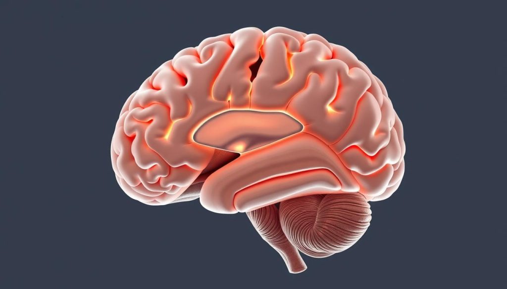
The calcarine sulcus is a key landmark on the brain’s medial surface. It’s linked to the visual cortex and contains the primary visual cortex (Brodmann area 17).
The calcarine sulcus, also known as the calcarine fissure, is a deep groove on the medial surface of the occipital lobe. It’s essential for processing visual information. Knowing the anatomy of the calcarine sulcus is vital in neuroanatomy, mainly when talking about the Angular Incisure.
The calcarine sulcus is a key part of the brain’s visual processing path. Its role and location are important for understanding many neurological conditions.
Anatomical Definition of the Angular Incisure
Anatomical studies have long focused on the angular incisure due to its clinical significance. It is a key landmark in the stomach’s anatomy. It plays a vital role in many physiological processes.
The angular incisure is found at the junction of the body and the pyloric part of the stomach. It has a sharp angular bend, making it easily recognizable.
Terminology and Classification
The terms used for the angular incisure come from its anatomical features. It is also called the incisura angularis. Anatomy classification systems group it based on its shape and structure.
| Term | Description |
|---|---|
| Incisura Angularis | A synonym for the angular incisure, stressing its angular shape. |
| Angular Incisure | The most common term, focusing on its location and shape. |
Historical Perspective in Anatomical Studies
Historically, the angular incisure has been of great interest. This is because of its role in gastric surgery and pathology. Early studies highlighted its importance in understanding stomach anatomy.
Today, with modern imaging, we have a better understanding of the angular incisure. This has helped us see its clinical significance in many gastrointestinal disorders.
Topographical Location of the Angular Incisure
Knowing where the angular incisure is located is key for both study and medical use. This important spot is on the stomach’s lesser curve. It’s where the stomach’s body meets the pyloric canal.
Precise Anatomical Coordinates
The angular incisure has a specific spot in the body. It’s near landmarks outside the body. Precise anatomical coordinates are vital for doctors to see on scans and during surgery. It’s near the lesser omentum, a part of the peritoneum that links the liver to the stomach and duodenum.
- It’s usually near the first or second lumbar vertebra.
- Its exact spot can change a bit from person to person.
- Scans like CT or MRI can find the angular incisure very accurately.
Relationship to Adjacent Structures
The angular incisure is near other important parts of the stomach. Knowing this helps doctors understand stomach problems. Its close relationship to the lesser omentum and the gastroesophageal junction is key.
Here’s how the angular incisure relates to nearby structures:
- It’s next to the lesser omentum, which has vital blood vessels and nerves.
- It’s close to the gastroesophageal junction, important for diagnosing GERD.
- Knowing its spot relative to the pyloric canal is key for surgeries in this area.
Structural Composition and Histology
The angular incisure is a key part of the digestive system. It has a special structure that helps it work well. Its layers and cells work together to keep it strong and functional.
Tissue Layers and Cellular Organization
The angular incisure has several layers. These include the mucosa, submucosa, muscularis externa, and serosa. The mucosa is the innermost layer. It has cells that help with absorption and secretion.
The submucosa is under the mucosa. It has blood vessels and nerves that support the mucosa. The muscularis externa is made of smooth muscle cells. These cells help the digestive tract move.
| Tissue Layer | Composition | Function |
|---|---|---|
| Mucosa | Epithelial cells | Absorption and secretion |
| Submucosa | Blood vessels and nerve fibers | Supports mucosa |
| Muscularis Externa | Smooth muscle cells | Motility |
Microscopic Features
The angular incisure has unique microscopic features. The mucosa has goblet cells that make mucus. This mucus helps food move smoothly.
The lamina propria in the mucosa has immune cells. These cells help fight off infections in the gut. Knowing about these features helps us understand the angular incisure’s role in health and disease.
Vascular and Neural Supply
Understanding the vascular and neural supply of the angular incisure is key. It shows its importance, mainly in abdominal surgery.
Arterial and Venous Networks
The angular incisure gets its blood from a network of arteries. These arteries branch off from the gastric arteries. The arterial supply is vital for the area’s health and function.
The venous drainage works with the arterial supply. This vascular network is essential for digestion.
Innervation Patterns
The innervation of the angular incisure is complex. It involves both sympathetic and parasympathetic nervous systems. The parasympathetic innervation, mainly from the vagus nerve, controls stomach functions.
The sympathetic innervation helps regulate blood flow. It also balances the parasympathetic effects.
This balance is key for the angular incisure’s proper function. It affects the whole gastrointestinal tract.
Relationship to the Lesser Omentum
The angular incisure’s connection to the lesser omentum is key in understanding the human body. Even though the calcarine sulcus is in the brain, knowing how different parts of the body link up is vital. This knowledge helps us grasp the full picture of human anatomy.
Anatomical Connections and Boundaries
The lesser omentum is a double-layered peritoneal fold that links the liver to the stomach and the start of the duodenum. The angular incisure, being part of the stomach, is closely tied to the lesser omentum. The lesser omentum’s free edge holds the portal vein, hepatic artery, and bile duct. These are essential for the liver’s work and are linked to the stomach’s structure.
The lesser omentum’s edges are marked by the hepatogastric and hepatoduodenal ligaments. These ligaments offer structural support and house vital vessels and ducts. They are key to the digestive process.
Functional Interrelationships
The angular incisure and the lesser omentum work together in gastrointestinal motility and secretion. The angular incisure helps break down food mechanically. The lesser omentum, connected to the liver and stomach, supports digestion by housing important structures.
The gastroesophageal junction, related to the angular incisure, is also affected by the lesser omentum’s arrangement. This junction is vital for stopping reflux and ensuring food moves properly into the stomach.
The Gastroesophageal Junction and Angular Incisure
The angular incisure and gastroesophageal junction are key parts of our digestive system. The gastroesophageal junction is where the esophagus meets the stomach. It’s important for stopping stomach acid from flowing back up. The angular incisure is a spot on the stomach’s lesser curvature.
Knowing how these parts work together helps doctors find and treat stomach problems.
Structural Continuity
The gastroesophageal junction and angular incisure are connected by muscles and mucous membranes. The lower esophageal sphincter at the junction stops stomach acid from coming back up. The angular incisure is close to the stomach’s lesser curvature.
Keeping these areas healthy is key for our digestive system to work right.
The way these parts are connected affects how our upper digestive system moves food. Problems in this connection can cause hiatal hernias, where the stomach pushes into the chest.
Physiological Significance
The gastroesophageal junction and angular incisure are vital for digestion. The junction keeps stomach acid from flowing back up. The angular incisure helps us understand how the stomach works.
Together, they help our upper digestive system work well.
Understanding these parts is important for treating conditions like GERD. The angular incisure’s role is also key for surgeries in this area.
Embryological Development and Formation
Learning about the angular incisure’s origins is key to understanding its role in the body. It shows how it fits into the growth of the gut during early development.
Developmental Stages
The angular incisure forms through several important steps. First, the early gut starts to take shape in the first weeks of pregnancy. It then splits into parts that will become different parts of the gut.
The angular incisure becomes clear as the stomach and the start of the duodenum grow and take their final shapes.
- The gut splits into foregut, midgut, and hindgut.
- The stomach grows from the foregut, with the angular incisure at its end.
- Its growth involves cells multiplying and changing types.
Congenital Variations and Anomalies
Some babies are born with the angular incisure not quite right. This can happen if something goes wrong during its growth. These issues can affect how the stomach and duodenum work.
Congenital anomalies may include:
- Problems with the angular incisure’s shape or where it is.
- Other issues with the gut.
Knowing about these problems helps doctors find and treat related health issues.
Radiological Assessment and Imaging Techniques
Advanced imaging has changed how we look at the angular incisure in medicine. These methods let doctors see the angular incisure clearly. This helps them make accurate diagnoses and plan treatments well.
Contrast Studies and Endoscopic Visualization
Contrast studies, like barium swallows, are great for checking the angular incisure’s shape and how it works. They spot problems like tight spots or odd shapes in the area.
Endoscopy gives a close-up look at the angular incisure. It checks the lining of the area and finds any issues or growths. It’s good for seeing how the angular incisure connects with other parts.
- Contrast studies help find structural issues.
- Endoscopy checks the lining’s health.
Cross-sectional Imaging Modalities
Imaging methods like CT and MRI scans show the angular incisure and what’s around it in detail. These are key for checking the angular incisure’s health and its ties to nearby areas.
CT scans show the angular incisure’s layout well. MRI scans, on the other hand, highlight soft tissues better. This helps doctors spot problems in this area.
Key benefits of cross-sectional imaging include:
- Clear views of the anatomy.
- Better accuracy in diagnosis.
Using these imaging and assessment methods, doctors can fully understand the angular incisure’s role in health. This leads to better care for patients.
Clinical Significance in Abdominal Surgery
Knowing about the angular incisure is key for surgeons in abdominal surgery. It’s a vital landmark that helps them during different surgeries.
Surgical Landmarks and Approaches
The angular incisure is a major landmark in abdominal surgery. It helps surgeons find other important structures. Surgical approaches often start with the angular incisure. It’s close to the stomach and esophagus, making it key in surgeries there.
Surgeons use it to know how much to dissect and to avoid harming nearby structures. The exact location and anatomy of the angular incisure can differ from person to person. So, imaging before surgery and checking it during surgery is very important.
Intraoperative Considerations
When doing abdominal surgery, the angular incisure is important for looking at the gastroesophageal junction’s anatomy. It’s important to keep the angular incisure intact to keep things working right. Surgeons need to know about the possible variations in the angular incisure’s anatomy and plan their surgery based on that.
The angular incisure’s role in abdominal surgery is big. It’s important for both finding landmarks and making decisions during surgery. By understanding the angular incisure, surgeons can do better in abdominal surgery.
Pathological Conditions Involving the Angular Incisure
The angular incisure is a key part of our body. It can get sick, which affects our stomach health.
Inflammatory and Neoplastic Disorders
Gastritis can harm the angular incisure, causing ulcers. Gastric cancer can also hit this area. Knowing about these problems helps doctors treat them better.
Inflammatory Conditions: Gastritis and ulcers are common problems. They often come from Helicobacter pylori or NSAIDs.
Diagnostic Criteria and Evaluation
To find out what’s wrong, doctors use several methods. They look inside with endoscopy and use CT scans to see how far the disease has spread.
| Diagnostic Method | Description | Clinical Utility |
|---|---|---|
| Endoscopy | Direct visualization of the gastric mucosa | Highly effective for diagnosing mucosal lesions and ulcers |
| CT Scan | Cross-sectional imaging of the abdomen | Useful for assessing the extent of disease and detecting masses |
| Biopsy | Histological examination of tissue samples | Critical for diagnosing neoplastic conditions |
It’s very important to diagnose and treat problems with the angular incisure right. This helps avoid serious issues and makes patients feel better.
Hiatal Hernia: Relationship with the Angular Incisure
Hiatal hernia happens when part of the stomach bulges into the chest. This can affect the area where the stomach meets the esophagus and the angular incisure.
The angular incisure is important because it’s close to where the stomach meets the esophagus. Knowing how hiatal hernia and the angular incisure are connected helps us understand the condition better.
Pathophysiological Mechanisms
Hiatal hernia develops due to several factors. These include increased intra-abdominal pressure and weakening of the diaphragmatic crura. These can cause the stomach to bulge into the chest, affecting the angular incisure.
- Increased intra-abdominal pressure
- Weakening of the diaphragmatic crura
- Disruption of the gastroesophageal junction
Clinical Manifestations and Management
Hiatal hernia can cause symptoms like heartburn, regurgitation, and dysphagia. Treatment options include lifestyle changes and medicines. In severe cases, surgery may be needed.
- Medical management: Lifestyle modifications and medications to reduce symptoms
- Surgical intervention: Procedures like fundoplication to repair the hernia
In summary, the connection between hiatal hernia and the angular incisure is key. Effective treatment involves understanding the condition’s causes and symptoms.
Functional Role in Digestive Physiology
Understanding the angular incisure is key to knowing its role in digestion. This special spot in the body plays a big part in how we digest food.
The angular incisure helps with many steps in digestion. It affects how food moves and how it’s broken down. Its role is important for digestion to work right.
Mechanical Functions and Motility
The angular incisure is important for digestion’s mechanical steps. It helps move food through the stomach and digestive system. It helps break down food with muscle contractions. This makes sure food mixes well with digestive enzymes.
Contribution to Digestive Processes
The angular incisure also helps with digestion in other ways. It controls when food moves from the stomach to the small intestine. This ensures nutrients are absorbed well. It keeps the stomach and esophagus working right, preventing problems like reflux.
Recent Advances in Understanding the Angular Incisure
Recent studies have greatly improved our knowledge of the angular incisure. This area is key to our health and has seen more research. It’s important for our digestive system and can help in diagnosing diseases.
Today’s research uses new imaging and tools to study the angular incisure. These tools help us understand its structure and function. They also show how it works with other parts of our body and aids in digestion.
Contemporary Research Findings
Recent studies show the angular incisure’s big role in medicine. They found that problems here can lead to many digestive issues. This makes it critical to diagnose and treat these issues correctly.
- Advanced imaging lets us see the angular incisure better, helping catch problems early.
- Research has also shown how the angular incisure is linked to some digestive diseases.
Emerging Concepts and Technologies
New ideas and technologies are helping us learn more about the angular incisure. Better endoscopy and imaging are giving doctors new ways to diagnose and treat. This is a big step forward in managing health issues related to the angular incisure.
- High-resolution endoscopy makes it easier to examine the angular incisure.
- CT and MRI scans are used to study the angular incisure in different health situations.
Using these new technologies in medicine will likely lead to better care. It will help doctors diagnose and treat problems with the angular incisure sooner and more effectively.
Conclusion
The Angular Incisure is a key part of the stomach’s anatomy. It’s important for understanding stomach health and diseases. We’ve looked at what it is, where it is, and why it matters.
The Angular Incisure’s anatomy is tied to the stomach’s structure and how it works. Knowing about its connection to nearby parts is key. This helps us see its importance in medical care.
In short, the Angular Incisure is a vital landmark in medicine. Studying it helps us learn more about the stomach and improve care. By understanding it, doctors can better treat patients and improve health outcomes.


