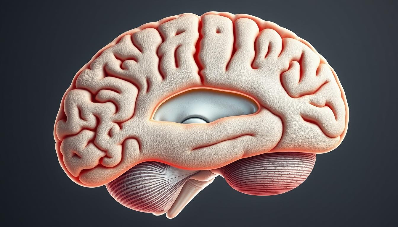
The calcarine sulcus is a key part of the brain’s surface. It’s found on the medial side of the brain. It’s important for how we see things.
Knowing about the anatomy of the calcarine sulcus helps us understand how we see. It’s where the primary visual cortex is. This makes it key to how we perceive what we see.
Studying the calcarine sulcus helps us learn more about how we see. It’s a big part of understanding human vision.
Overview of the Calcarine Sulcus
Understanding the calcarine sulcus is key to grasping how we see. It’s a major landmark in the occipital lobe. It plays a big role in how our visual cortex is organized.
Definition and Basic Characteristics
Anatomical Definition
The calcarine sulcus is a groove on the medial surface of the occipital lobe. It stretches from the occipital pole to the parieto-occipital sulcus. This complex structure is vital for our visual pathway to work right.
Gross Appearance
The calcarine sulcus looks like a deep, complex groove. It starts near the occipital pole and goes forward to a spot below the splenium of the corpus callosum. Its anatomy is linked to how our visual system functions.
Key Features of the Calcarine Sulcus:
- Location on the medial surface of the occipital lobe
- Extension from the occipital pole to the parieto-occipital sulcus
- Complex and deep structure
The calcarine sulcus is a vital part of brain anatomy, focusing on visual processing. Its unique structure and location make it a key area for neuroanatomy study.
Anatomical Location and Structure
The calcarine sulcus is on the medial surface of the occipital lobe. It’s key for processing visual information. It’s a major landmark in the brain’s visual pathway.
Position Within the Occipital Lobe
The calcarine sulcus is in the occipital lobe, on its medial side. It’s near the occipital pole. It divides the medial surface into the cuneus and the lingual gyrus. This division is vital for understanding the visual cortex’s function.
Relationship to Occipital Pole
The calcarine sulcus is close to the occipital pole. This closeness is important for its role in visual processing. Research shows it’s involved in early visual processing stages.
Medial Surface Orientation
The calcarine sulcus is on the medial surface of the occipital lobe. As Kenhub notes, “the calcarine sulcus is located on the medial surface of the occipital lobe.”
This orientation helps it divide the visual cortex into different areas
. Its medial surface orientation is key to its visual processing function.
The calcarine sulcus’s location and structure are key to its role in the visual pathway. Knowing its position in the occipital lobe and its relation to other structures is essential. It helps us understand its importance.
Embryological Development
Learning about the calcarine sulcus’s growth in the womb is key to understanding its adult form. This important part of our brain’s visual pathway starts forming in the fetus.
Fetal Development Timeline
The calcarine sulcus’s growth is a detailed process that spans several weeks in the womb. Major changes occur between the 20th and 28th weeks.
First Trimester Development
In the first trimester, the groundwork for the calcarine sulcus is set. It’s not seen yet, but the cells that will make up the sulcus start to organize.
Second and Third Trimester Maturation
By 20 to 28 weeks, the calcarine sulcus starts to show, as the brain grows a lot. This time is vital for its development. Any issues can cause problems.
Key milestones in the development include:
- The initial formation of the calcarine sulcus during the first trimester.
- Visible development between the 20th and 28th weeks of gestation.
- Continued maturation throughout the second and third trimesters.
The growth of the calcarine sulcus is tightly controlled. Any problems during key times can lead to big changes or issues.
Knowing this timeline helps us understand the brain’s complex adult structure. It also shows the importance of avoiding problems during development.
Microscopic Anatomy of the Calcarine Sulcus
Exploring the microscopic anatomy of the calcarine sulcus reveals its importance. It’s a key part of the visual pathway. The sulcus has a complex cellular structure.
Cellular Organization
The cellular organization in the calcarine sulcus is complex. It includes different neurons and glial cells. These cells work together to process visual information.
Neuronal Types and Distribution
The calcarine sulcus has many neuronal types. These include stellate, pyramidal, and basket cells. Each type has its own role in visual processing.
Glial Components
The sulcus also has glial components like astrocytes and oligodendrocytes. These cells support neurons and help signal transmission.
The interaction between neuronal types and glial components is key. The microscopic anatomy of the calcarine sulcus shows how visual info is processed.
Calcarine Sulcus Vascular Supply
The calcarine sulcus gets its blood from the posterior cerebral artery. This area is key for seeing things. It needs a special blood network to work right. This network comes from the calcarine artery, a part of the posterior cerebral artery.
Arterial Supply
The main blood source for the calcarine sulcus is the calcarine artery. It comes from the posterior cerebral artery. This artery is very important for the visual cortex, showing how vital the posterior cerebral artery is for seeing.
Posterior Cerebral Artery Distribution
The posterior cerebral artery feeds the occipital lobe, where the calcarine sulcus is. It has branches that help the visual cortex work. The calcarine artery, a part of this, makes sure the calcarine sulcus gets enough blood.
Collateral Circulation
The calcarine sulcus also has a backup blood supply. This is important when the main arteries get blocked. This backup helps keep the visual cortex working, even when things go wrong.
The blood flow to the calcarine sulcus is complex. It involves both main and backup supplies. Knowing about this is key for treating vision problems.
The role of the posterior cerebral artery and its branches is huge. It’s very important for the calcarine sulcus. Knowing about this helps doctors understand and treat vision issues better.
- The calcarine sulcus gets its main blood from the calcarine artery.
- The calcarine artery comes from the posterior cerebral artery.
- There’s also a backup blood supply.
Doctors can better handle vision problems by knowing about the blood flow to the calcarine sulcus.
Functional Neuroanatomy
Understanding the calcarine sulcus is key to knowing how we see. It’s where the primary visual cortex is, which starts to process what we see.
Role in Visual Processing
The calcarine sulcus is vital for visual processing. It’s home to the primary visual cortex. This part of the brain figures out basic things like lines, where things are, and if they’re moving.
Primary Visual Functions
The primary visual cortex in the calcarine sulcus deals with basic visual stuff. It spots line orientation, spatial location, and movement. This is the first step in making sense of what we see.
Integration with Visual Association Areas
After the primary visual cortex, the brain mixes this info with other visual association areas. These areas help with more complex tasks like recognizing objects and understanding space.
This mixing of information is key to seeing and understanding things. The calcarine sulcus, with its primary visual cortex, is at the heart of this process.
| Visual Processing Stage | Function | Associated Brain Area |
|---|---|---|
| Primary Processing | Line orientation, spatial location, movement | Primary Visual Cortex (Calcarine Sulcus) |
| Secondary Processing | Object recognition, spatial awareness | Visual Association Areas |
The table shows the different parts of seeing and where they happen in the brain. The primary visual cortex, in the calcarine sulcus, is where it all starts.
The Primary Visual Cortex and Calcarine Sulcus
The primary visual cortex, also known as V1, is closely tied to the calcarine sulcus. It plays a key role in processing visual information. The calcarine sulcus is a deep groove in the occipital lobe where V1 is found.
The primary visual cortex is structured as the striate cortex. This structure is vital for its role in visual processing.
Striate Cortex Organization
The striate cortex has a unique cellular layout. This layout is essential for processing visual information.
Cellular Architecture of V1
V1’s cellular structure includes various layers and cell types. These layers are arranged to handle different parts of visual stimuli. The cellular organization helps in efficiently passing on visual information.
Functional Columns
The primary visual cortex is divided into functional columns. These columns handle different aspects of visual information. They are key for analyzing visual stimuli, allowing the brain to understand complex visual data.
Studies have found that these columns are vital for processing visual attributes like orientation and spatial frequency (Source: StatPearls).
| Layer | Cell Type | Function |
|---|---|---|
| Layer 4 | Stellate cells | Receives input from the lateral geniculate nucleus |
| Layer 2/3 | Pyramidal cells | Processes visual information and sends output to higher visual areas |
| Layer 6 | Pyramidal cells | Sends feedback to the lateral geniculate nucleus |
The primary visual cortex’s organization in the calcarine sulcus is complex and specialized. Understanding this is key to grasping how visual information is processed.
Neuroimaging of the Calcarine Sulcus
Researchers can now explore the calcarine sulcus in new ways thanks to neuroimaging. This field is key to understanding brain anatomy and function. It helps us see how the brain processes visual information.
MRI Visualization Techniques
Magnetic Resonance Imaging (MRI) is a powerful tool for brain scans. It shows the calcarine sulcus and its surroundings in detail. MRI images are essential for both research and medical diagnosis.
Structural MRI Approaches
Structural MRI is great for looking at the calcarine sulcus’s anatomy. It gives clear images of the brain’s structure. This helps researchers study the sulcus’s shape and how it varies among people. Structural MRI has greatly helped us understand brain anatomy.
Diffusion Tensor Imaging Applications
Diffusion Tensor Imaging (DTI) is another important tool. It shows the white matter tracts around the calcarine sulcus. DTI tracks water diffusion in the brain, revealing nerve fiber details. This is key for studying visual pathway connections.
Using both structural MRI and DTI gives a full picture of the calcarine sulcus. These methods are vital in neuroscience, giving us deep insights into brain structure and function.
- Neuroimaging techniques enhance our understanding of the calcarine sulcus.
- MRI is key for seeing the calcarine sulcus’s anatomy.
- DTI sheds light on visual pathway connections.
In conclusion, MRI and related techniques have greatly advanced neuroscience. They provide detailed views of the calcarine sulcus. These breakthroughs are significant for both research and medical use.
Clinical Significance of the Calcarine Sulcus
Understanding the calcarine sulcus is key for diagnosing and treating visual problems. This area is vital for processing what we see.
Lesions and Associated Visual Field Defects
Damage to the calcarine sulcus can lead to specific vision problems. The type and severity of these issues depend on where and how severe the damage is.
Homonymous Hemianopia
Homonymous hemianopia means losing half of your vision in both eyes. Damage to the calcarine sulcus can cause this, greatly affecting your vision.
Quadrantanopia
Quadrantanopia is losing a quarter of your vision. It happens when a part of the calcarine sulcus is damaged. This shows how important this area is for seeing.
| Visual Field Defect | Description | Common Cause |
|---|---|---|
| Homonymous Hemianopia | Half-vision loss on the same side in both eyes | Lesions in the calcarine sulcus or optic tract |
| Quadrantanopia | Quarter vision loss | Damage to the calcarine sulcus or visual pathway |
The calcarine sulcus is very important because of the different vision problems it can cause. Proper diagnosis and treatment are essential for helping patients.
Pathological Conditions Affecting the Calcarine Sulcus
The calcarine sulcus is key for our vision. It can be damaged by stroke and ischemia. This damage can cause big problems with our sight.
Stroke and Ischemic Events
Stroke and ischemic events often harm the calcarine sulcus. The posterior cerebral artery feeds this area. When it gets blocked, the sulcus can be damaged.
Posterior Cerebral Artery Occlusion
When the posterior cerebral artery is blocked, it can cause vision problems. This is because the calcarine sulcus gets its blood from this artery.
Clinical Presentation and Management
People with blocked posterior cerebral artery might lose half of their vision. They might see less on the same side in both eyes. Doctors treat this with acute stroke care and managing stroke risks.
Clinical Features and Management of Posterior Cerebral Artery Occlusion
| Clinical Features | Management Strategies |
|---|---|
| Homonymous hemianopia | Acute stroke care, including thrombolysis |
| Visual field defects | Rehabilitation and visual therapy |
| Potential cognitive impairments | Cognitive rehabilitation and management of risk factors |
In summary, stroke and ischemia can harm the calcarine sulcus, leading to vision problems. Knowing how to treat these conditions is key to helping patients.
Comparative Anatomy Across Species
The calcarine sulcus is not just found in humans. It’s also present in other primates. This shows its key role in how we see the world. It helps process visual information, and its presence in different species shows it’s an important part of evolution.
Evolutionary Perspectives
The calcarine sulcus has a big role in evolution. It’s found in many primates, showing they share a common ancestor. This shared feature is important for understanding how vision evolved.
Phylogenetic Development
The calcarine sulcus’s development is tied to the evolution of vision in primates. Research shows it’s very similar across species. This means it’s been essential for vision for millions of years.
Functional Adaptations
The calcarine sulcus helps process complex visual information. Its ability to handle complex sights has helped it stay the same over time. It plays a big part in how primates see and interact with their world.
In summary, the calcarine sulcus is very important in evolution and vision. By studying how it has changed and stayed the same, we learn a lot about how vision evolved in primates.
Modern Research and Discoveries
Modern research is revealing the details of the calcarine sulcus and its role in vision. Advances in neuroimaging and studies have greatly improved our knowledge of this key brain area.
Recent Scientific Findings
Recent studies have looked into the neuroplasticity and functional connectivity of the calcarine sulcus. They’ve found new insights into its ability to adapt and work with other brain parts. Research in StatPearls shows the calcarine sulcus can change a lot in response to different visual inputs.
Neuroplasticity Studies
Studies on neuroplasticity show the calcarine sulcus can change itself after injury or when visual input changes. This reorganizational capability is key for understanding how patients with vision problems recover.
Functional Connectivity Research
Research on functional connectivity has shown the calcarine sulcus’s detailed connections with other visual areas. It plays a big role in the visual pathway, helping with complex visual tasks.
| Research Focus | Key Findings |
|---|---|
| Neuroplasticity | The calcarine sulcus shows significant adaptability in response to visual changes or injury. |
| Functional Connectivity | The calcarine sulcus is highly connected with other visual processing areas, facilitating complex visual tasks. |
The latest research on the calcarine sulcus is changing how we treat and understand vision. As we learn more about this brain area, new ways to help people with vision problems are being found.
Surgical Considerations
Surgeries in the calcarine sulcus need careful planning to keep vision intact. This area’s complexity requires a deep understanding of its anatomy and the risks of surgery.
Neurosurgical Approaches
Neurosurgery in the calcarine sulcus is complex. It requires precise methods to reach the occipital lobe without harming nearby areas. Preoperative planning is key to find the best surgical path.
Occipital Lobe Access Techniques
There are different ways to reach the occipital lobe, like lateral and medial approaches. The choice depends on the patient’s condition and anatomy.
Intraoperative Monitoring
Monitoring during surgery is vital to protect vision in the calcarine sulcus. Visual evoked potentials (VEPs) help track the visual pathway in real-time.
Studies show that monitoring during surgery improves results near the calcarine sulcus.
A team of neurosurgeons, neurologists, and radiologists is needed for calcarine sulcus surgeries. Advanced techniques, careful planning, and monitoring help reduce surgery risks in this sensitive area.
Conclusion
The calcarine sulcus is key in visual processing. Knowing about it is important for doctors and researchers. Its detailed brain anatomy helps us understand clinical data better.
Damage to the calcarine sulcus can cause big problems with seeing. This shows how important it is. Thanks to new tech, we can now better diagnose and treat these issues.
Understanding the calcarine sulcus is a must for doctors and scientists. More research will help us learn more about its role in visual processing. This knowledge is vital for understanding and treating brain disorders.