The Dentoalveolar Joint is key to keeping teeth in line and working right. It’s a big part of Dental Anatomy and Oral Health.
This complex part, also called the tooth socket or alveolar bone, is vital. It supports the teeth and helps with many oral functions.
Knowing about the Dentoalveolar Joint helps us see its importance in dental health. It also helps us spot problems that could affect our mouth’s health.
The Dentoalveolar Joint: Definition and Overview
Understanding the Dentoalveolar Joint is key to knowing its role in eating and speaking. This joint, also known as the tooth socket, holds the tooth in place with the alveolar bone.
Anatomical Classification and Terminology
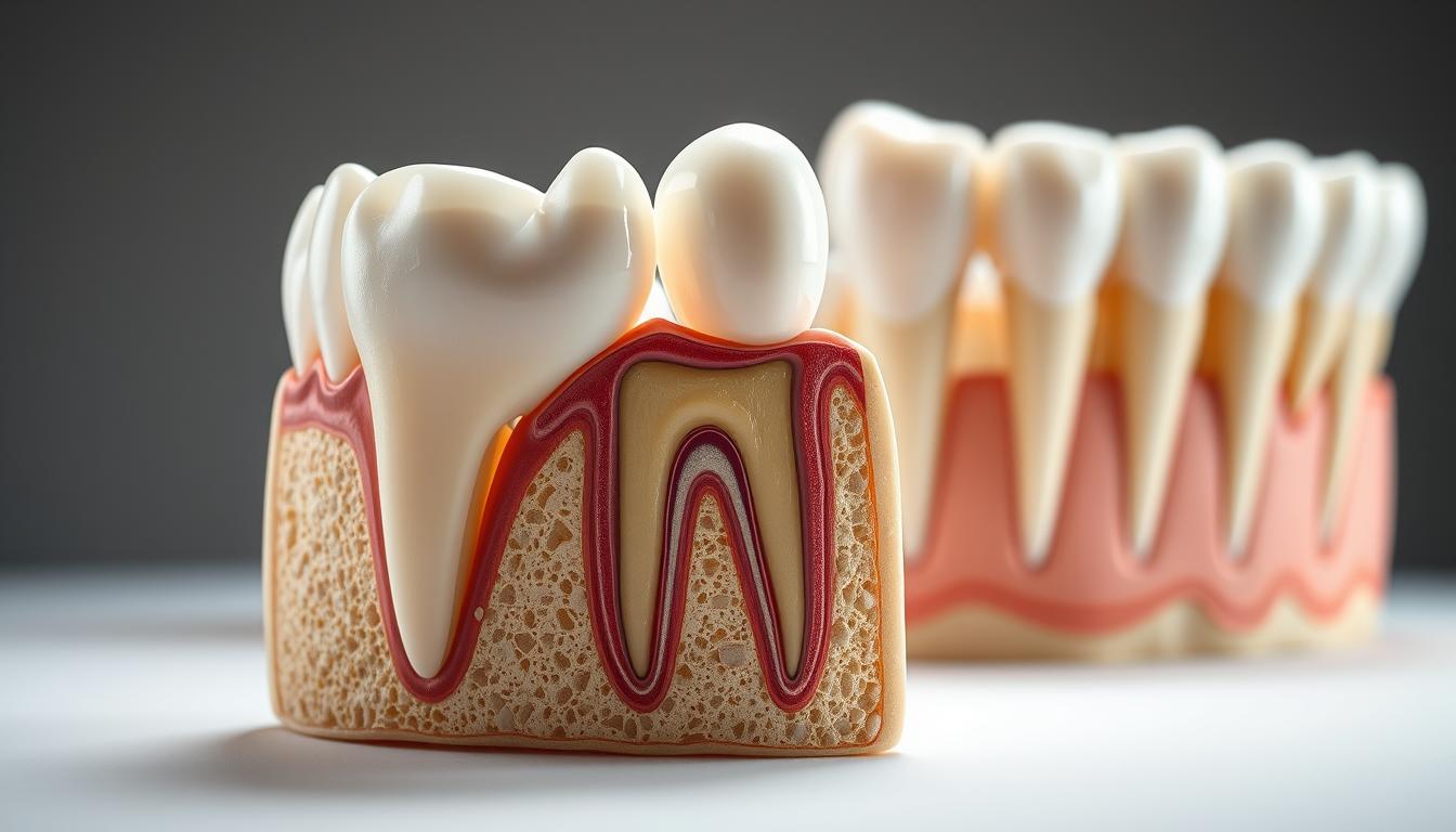
The Dentoalveolar Joint is a type of joint that doesn’t move. It’s a fibrous joint that links the tooth to the alveolar bone through the periodontal ligament. The terms related to this joint include the tooth, alveolar bone, and periodontal ligament.
Key parts of the Dentoalveolar Joint are:
- The tooth root and its shape
- The alveolar bone that holds the tooth socket
- The periodontal ligament that ties the tooth to the alveolar bone
Evolutionary Development and Significance
The Dentoalveolar Joint evolved to help with eating, speaking, and swallowing. Its growth is tied to the development of teeth and alveolar bone. This joint is important because it helps absorb forces during chewing, protecting the tooth and bone.
The development of the Dentoalveolar Joint is a complex process. It involves the growth of the tooth, periodontal ligament, and alveolar bone together. This joint is vital for keeping the mouth healthy and working well.
Structural Components of the Dentoalveolar Joint
It’s important to know about the dentoalveolar joint’s parts to understand its role. This joint is key in the mouth, helping with chewing, talking, and swallowing.
The dentoalveolar joint has several important parts that work together for oral health. These include the tooth, alveolar bone, and periodontal ligament. Each part has a special role in how the joint works.
Tooth Structure and Root Morphology
The tooth has a unique shape that affects how it fits with the bone and ligament. The shape of the root is important for how forces are spread in the tooth socket. Different shapes and sizes can change how stress is put on the alveolar bone.
Alveolar Bone Architecture
The alveolar bone supports the teeth and changes with the forces of chewing. Its structure and density are key for the joint’s stability and health.
Periodontal Ligament Organization
The periodontal ligament is a tissue that surrounds the tooth root and connects it to the alveolar bone. It helps distribute forces, sense movement, and keep the mouth healthy. The health of the periodontal ligament is essential for the joint’s proper function.
In summary, the parts of the dentoalveolar joint work together for its functions. Knowing about the tooth, alveolar bone, and periodontal ligament is key for good oral health and function.
Microscopic Anatomy of the Dentoalveolar Complex
The dentoalveolar complex is made up of cells, matrix, and blood vessels. This complex structure is key to its function and health.
Cellular Components and Their Functions
The dentoalveolar complex has different cell types, each with its own job. Osteoblasts and osteoclasts help make and break down bone. They are important for bone growth and repair.
Fibroblasts in the periodontal ligament make and keep the matrix. Cementoblasts and cementoclasts work on cementum, a special bone-like layer on the tooth root. These cells work together to keep the dentoalveolar complex strong and functional.
Extracellular Matrix Composition
The extracellular matrix is vital for the dentoalveolar complex. It gives structure and helps cells talk to each other. The matrix in the periodontal ligament has collagen fibers, which make it strong and flexible.
Vascular and Neural Supply
The dentoalveolar complex gets a lot of blood, which it needs to live and work. Blood brings oxygen and nutrients to the cells, including those in the periodontal ligament and bone.
The nerves in the dentoalveolar complex are also important. They help with feeling and sensing. The nerves in the periodontal ligament are key for feeling pressure and force, helping with chewing and biting.
| Component | Function | Key Cells Involved |
|---|---|---|
| Alveolar Bone | Supports tooth anchorage | Osteoblasts, Osteoclasts |
| Periodontal Ligament | Attaches tooth to bone, shock absorption | Fibroblasts |
| Cementum | Tooth root covering, attachment for PDL | Cementoblasts, Cementoclasts |
Comparative Anatomy of the Dentoalveolar Joint
The dentoalveolar joint changes a lot in different mammals. This shows how it adapts to different diets and environments. It’s key for various eating and oral functions.
Variations Across Mammalian Species
Studies reveal the dentoalveolar joint’s shape changes based on diet, jaw use, and evolution. Herbivores have a more complex joint than carnivores. This is because of the different forces their diets put on their jaws.
Functional Adaptations in Different Dentition Types
The joint also changes based on tooth types. For example, carnivores with short teeth have a unique joint. Herbivores with long teeth have a different one. These changes help the joint handle the specific needs of each tooth type.
In summary, the dentoalveolar joint’s anatomy shows its amazing ability to adapt. It’s vital for the health and function of different mammals’ mouths.
Functional Biomechanics of the Dentoalveolar Joint
It’s key to know how the dentoalveolar joint works for chewing and biting. This joint, made of tooth, periodontal ligament, and alveolar bone, acts as a single unit.
Force Distribution During Mastication and Occlusion
When we chew, the dentoalveolar joint faces different forces. The periodontal ligament helps spread these forces, keeping the alveolar bone and tooth safe.
The strength and direction of these forces matter a lot. Good biting alignment is vital for the joint’s health. Bad alignment can cause uneven force and harm.
Adaptive Remodeling Mechanisms
The dentoalveolar joint can change to handle new forces. The alveolar bone is always being remodeled by osteoblasts and osteoclasts.
This ability is key for adjusting to changes in biting forces and keeping the joint healthy. The periodontal ligament also changes with orthodontic forces, allowing teeth to move.
Proprioceptive Feedback Systems
Inside the dentoalveolar joint, there are systems that send feedback on its position and forces. The periodontal ligament has sensors that feel changes in force and send signals to the brain.
This feedback is vital for controlling chewing forces and keeping biting in balance. It helps control jaw movements, making sure we chew safely and efficiently.
Embryological Development of the Dentoalveolar Joint
Understanding how the dentoalveolar joint forms is key to knowing its role. This process starts early in a fetus’s development. It involves many complex steps.
Early Formation and Differentiation
The journey begins with the ectodermal layer, which becomes the dental epithelium. This layer works with the mesenchyme below. Together, they create the cells needed for teeth and bone.
Cell differentiation is vital. It leads to the creation of different cell types. These include odontoblasts, cementoblasts, and osteoblasts.
Stages of Tooth and Alveolar Development
Tooth growth goes through several stages: initiation, proliferation, histodifferentiation, and morphodifferentiation. Each stage is essential for the tooth’s proper formation.
- Initiation: The dental lamina forms, starting tooth development.
- Proliferation: The dental epithelium grows, forming the tooth bud.
- Histodifferentiation: Cells become specific types, like ameloblasts and odontoblasts.
- Morphodifferentiation: The tooth’s final shape is determined.
At the same time, the alveolar bone develops around the tooth. This bone provides the necessary support.
Molecular Signaling in Joint Formation
Molecular signals are critical in forming the dentoalveolar joint. Growth factors and signaling molecules, like BMPs and Wnts, control cell growth, differentiation, and survival.
| Signaling Molecule | Function |
|---|---|
| BMPs | Regulate osteoblast differentiation and bone formation |
| Wnts | Influence cell fate decisions and tissue patterning |
The complex interaction of these signals is what ensures the dentoalveolar joint forms and functions correctly.
The Role of the Periodontal Ligament in Joint Function
The periodontal ligament is key to the dentoalveolar joint’s success. It connects the tooth to the bone around it. This connection is vital for the joint’s health and function.
Shock Absorption and Force Distribution
The periodontal ligament helps absorb shock and distribute forces during chewing. Its structure and cells work together to protect the tooth from too much stress.
This protection is essential for the tooth and bone’s health. It helps the dentoalveolar joint last longer. The ligament’s ability to handle different forces shows its importance in keeping the mouth healthy.
Sensory Function and Mechanoreception
The periodontal ligament also has sensory roles. It contains mechanoreceptors that send signals about tooth forces. This feedback is key for controlling chewing and other mouth movements.
These mechanoreceptors are very sensitive to pressure and tension. They help control how we move our mouth. This feedback loop is vital for the joint’s health and function.
Nutritive, Reparative, and Homeostatic Roles
The periodontal ligament also helps keep the surrounding tissues healthy. It provides cells and nutrients for repairing and maintaining the bone and other tissues.
| Function | Description |
|---|---|
| Shock Absorption | Absorbs forces during mastication, protecting the tooth and surrounding bone. |
| Sensory Function | Provides feedback on forces applied to the tooth, regulating oral functions. |
| Nutritive and Reparative Roles | Supplies cells and nutrients for the repair and maintenance of periodontal tissues. |
In conclusion, the periodontal ligament is essential for the dentoalveolar joint’s health. Its roles in shock absorption, sensory feedback, and nutrition are vital. These functions ensure the joint’s longevity and efficiency.
Alveolar Bone Physiology and Remodeling
Understanding alveolar bone remodeling is key to dental anatomy and oral health. The alveolar bone is dynamic, supporting teeth and constantly remodeling.
Bone Cells: Osteoblasts, Osteoclasts, and Osteocytes
The alveolar bone has different cells like osteoblasts, osteoclasts, and osteocytes. Osteoblasts form bone, making the matrix and controlling mineralization. Osteoclasts break down bone tissue. Osteocytes are mature cells in the bone matrix, important for sensing and regulating bone remodeling.
The Remodeling Cycle and Regulation
The alveolar bone remodeling cycle involves osteoblasts and osteoclasts working together. This process is controlled by hormones, growth factors, and mechanical loading.
| Cell Type | Function |
|---|---|
| Osteoblasts | Bone formation |
| Osteoclasts | Bone resorption |
| Osteocytes | Mechanosensing and regulation |
Systemic and Local Factors Affecting Bone Metabolism
Systemic and local factors impact alveolar bone metabolism. Hormones like parathyroid hormone and estrogen are systemic factors. Local factors include mechanical loading and inflammatory mediators.
In conclusion, alveolar bone physiology and remodeling are complex. They are vital for oral health. Understanding these processes helps in diagnosing and treating dental disorders.
Pathological Conditions Affecting the Dentoalveolar Joint
It’s important to know about the diseases that can harm the dentoalveolar joint. This joint is complex and can get sick in many ways. This can hurt its function and overall health.
Periodontal Disease: Pathogenesis and Progression
Periodontal disease is a big problem for the dentoalveolar joint. It causes inflammation and can damage the teeth’s support. The disease starts with plaque, the body’s immune response, and other factors.
Key stages in the progression of periodontal disease include:
- Gingivitis, characterized by inflammation of the gingiva
- Periodontitis, involving destruction of the periodontal ligament and alveolar bone
- Advanced periodontitis, leading to significant loss of tooth support
Traumatic Injuries and Fracture Patterns
Trauma can damage the dentoalveolar joint, affecting its health. The type of fracture depends on the force and direction of the injury.
| Type of Fracture | Description | Common Causes |
|---|---|---|
| Tooth Fracture | Fracture of the tooth structure | Trauma, biting on hard objects |
| Alveolar Bone Fracture | Fracture of the alveolar bone supporting the teeth | Severe trauma to the jaw |
Developmental and Congenital Anomalies
Birth defects can also harm the dentoalveolar joint. This includes conditions like ankylosed teeth and extra teeth.
Ankylosed Teeth and Fusion Disorders
Ankylosed teeth are stuck to the bone, causing problems with tooth growth and orthodontics. Fusion disorders join two or more teeth together.
Supernumerary Teeth and Positional Abnormalities
Extra teeth can cause crowding and other issues. These problems might need orthodontic or surgical fixes.
In conclusion, the dentoalveolar joint faces many health issues. These include periodontal disease, injuries, and birth defects. Knowing about these problems helps in treating them and keeping the mouth healthy.
Diagnostic Approaches for Dentoalveolar Joint Assessment
Assessing the dentoalveolar joint requires a mix of clinical checks and advanced imaging. This combo helps understand its structure and how it works.
Clinical Examination Techniques and Protocols
First, a detailed clinical check is done. This includes looking at the patient’s health history and dental records. It also involves a visual check of the mouth and tests to find any pain or issues.
These tests might also check how well the jaw moves and the alignment of teeth. They look for any signs of injury or disease.
Conventional and Digital Radiographic Evaluation
Imaging is key in diagnosing dentoalveolar joint problems. Traditional X-rays show the bone and teeth well.
Digital X-rays, on the other hand, give clearer images and use less radiation. They also let doctors adjust the images for better diagnosis.
| Radiographic Technique | Advantages | Limitations |
|---|---|---|
| Conventional Radiography | Wide availability, cost-effective | Limited detail, radiation exposure |
| Digital Radiography | Enhanced image quality, reduced radiation | Higher initial cost, technical requirements |
Advanced Imaging: CBCT, MRI, and Ultrasound Applications
Techniques like CBCT, MRI, and ultrasound give detailed views of the dentoalveolar joint. They help spot problems and understand the joint’s anatomy.
“CBCT has revolutionized the field of dental imaging by providing three-dimensional views of the dental structures, allowing for more accurate diagnoses and treatment plans.”
These tools are great for complex cases, like TMJ disorders or trauma. They help doctors plan better treatments.
In summary, a mix of clinical checks, X-rays, and advanced imaging is vital for a full dentoalveolar joint assessment. This approach helps dental experts make accurate diagnoses and treatments. It keeps our mouths healthy.
Therapeutic Interventions for Dentoalveolar Joint Disorders
Managing dentoalveolar joint disorders needs a mix of treatments. Each plan is made for the patient’s specific needs. This depends on the type and how severe the condition is.
Conservative Management Strategies
First, doctors often try conservative management. This might include:
- Occlusal adjustments to improve bite alignment
- Physical therapy to enhance jaw mobility and strength
- Pharmacological interventions for pain management
- Lifestyle modifications, such as dietary changes and stress reduction techniques
A leading expert says, “Conservative management can greatly help patients. It reduces pain and improves jaw function.”
“The goal of conservative management is to alleviate symptoms and improve jaw function without resorting to invasive procedures.”
Surgical Approaches and Techniques
If conservative methods don’t work, surgery might be needed. Surgical options include:
- Arthrocentesis to lavage and medicate the joint
- Arthroscopy for direct visualization and treatment of joint pathology
- Open-joint surgery for more complex conditions or when other treatments have failed
| Surgical Technique | Indications | Benefits |
|---|---|---|
| Arthrocentesis | Acute joint inflammation, pain | Reduces pain, improves mobility |
| Arthroscopy | Internal joint derangements | Direct visualization, minimally invasive |
| Open-joint surgery | Complex joint pathology, failed previous treatments | Allows for complete repair or reconstruction |
Regenerative Therapies and Tissue Engineering
Regenerative therapies are a new hope for treating dentoalveolar joint disorders. They include:
- Platelet-rich plasma (PRP) therapy to stimulate healing
- Stem cell therapy for tissue regeneration
- Tissue engineering approaches to repair or replace damaged tissues
These therapies aim for lasting results. They fix the root cause and help tissues heal.
The Dentoalveolar Joint in Orthodontic Treatment
The dentoalveolar joint changes in many ways when we get orthodontic treatment. This treatment uses forces to move teeth, affecting the joint and its tissues.
Biological Basis of Tooth Movement
Tooth movement in orthodontic treatment starts with the dentoalveolar joint’s response. Forces applied to teeth cause inflammation, bone breakdown, and new bone growth. The periodontal ligament is key, connecting the tooth to the bone.
The tooth movement process includes several important steps:
- Inflammation and cellular response
- Bone remodeling through resorption and formation
- Periodontal ligament adaptation
Tissue Response to Different Types of Orthodontic Forces
The dentoalveolar joint and tissues react differently to orthodontic forces. Knowing how they respond helps plan treatment better. The force’s type, strength, and how long it’s applied affects treatment success.
Here’s how different forces impact tissues:
- Continuous forces lead to ongoing bone remodeling.
- Intermittent forces cause partial bone remodeling.
- Pulsating forces boost cell activity.
Long-term Effects on Joint Integrity and Function
Orthodontic treatment’s long-term effects on the dentoalveolar joint are important. Good treatment leads to lasting results. But, bad or too much force can harm the joint and teeth.
To get the best results, treatment needs careful planning and watching. This includes:
- Checking tooth movement and joint response often
- Changing treatment forces when needed
- Keeping up with follow-ups for oral health
Conclusion: The Importance of Dentoalveolar Joint Health in Overall Oral Function
The Dentoalveolar Joint is key to our mouth’s health. It helps us chew, speak, and keep our mouth healthy. This joint’s complex structure and function show how important it is for our mouth.
It’s vital to take care of this joint to avoid problems and keep our mouth healthy. Knowing how the joint works and grows helps us understand its role in our mouth’s function.
Keeping the Dentoalveolar Joint healthy is essential for our mouth and overall health. By taking care of it, we can keep our mouth working well and avoid many health issues.