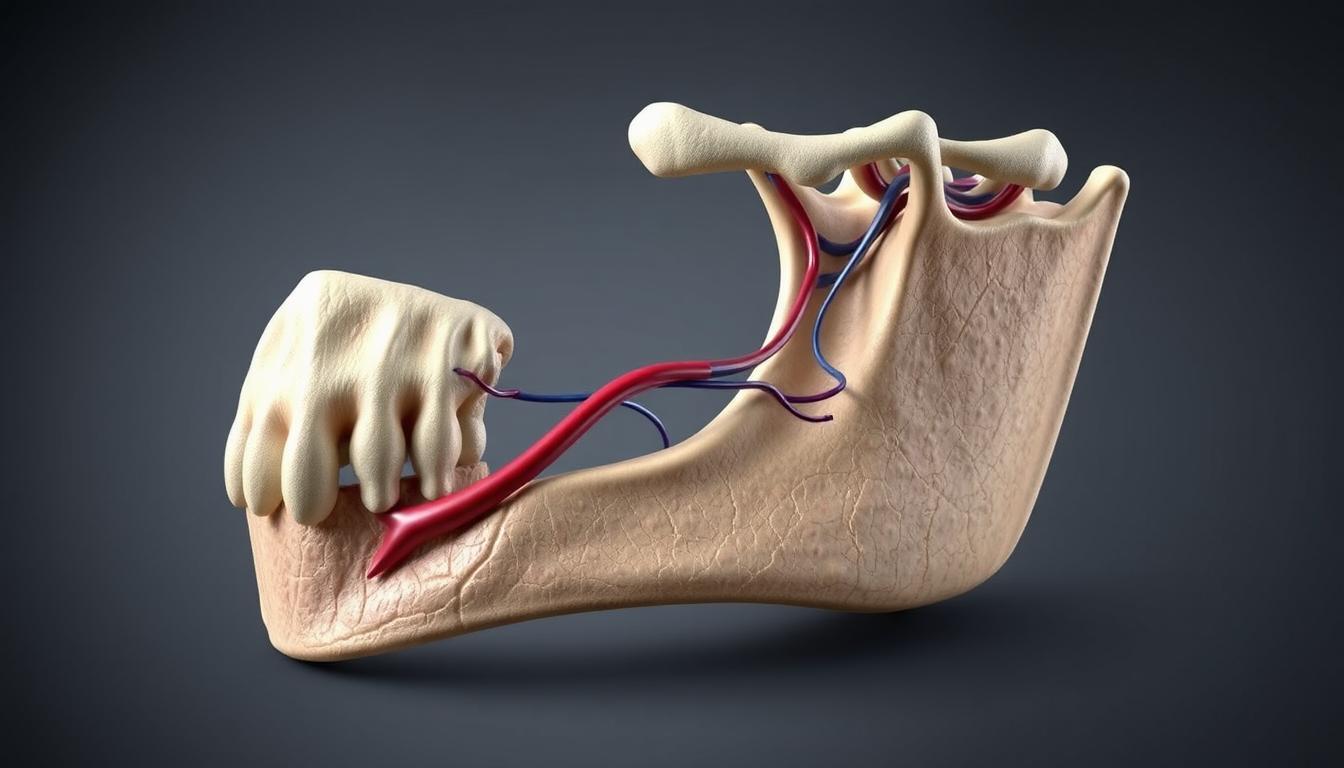
The retromandibular vein is a key deep vein in the face. It plays a big role in draining blood from the head and neck.
It forms when the superficial temporal vein meets the maxillary vein. This vein goes through the parotid gland. It then goes down behind the mandible’s ramus.
Knowing the anatomy of the retromandibular vein helps us understand the head and neck’s complex neuroanatomy and cranial nerves.
The vein’s location and how it’s formed show its importance in the venous drainage system. It’s a key part of the region’s anatomy.
Overview of the Retromandibular Vein
Understanding the retromandibular vein is key to knowing the head and neck’s venous system. This vein is vital for draining blood from the jaw, lateral skull, and parotid gland.
Definition and Basic Characteristics
The retromandibular vein is made by the joining of the superficial temporal and maxillary veins. It’s found in the parotid gland and is a major part of the venous drainage system in the head and neck.
The retromandibular vein is special because of its location and path. It goes through the parotid gland, next to the external carotid artery. It then empties into either the external or internal jugular vein.
| Characteristics | Description |
|---|---|
| Formation | Confluence of superficial temporal and maxillary veins |
| Location | Within the parotid gland |
| Course | Runs alongside the external carotid artery |
| Drainage | External or internal jugular vein |
Importance in Head and Neck Venous Drainage
The retromandibular vein is vital for draining blood from the head and neck. It takes blood from the jaw, lateral skull, and parotid gland. Then, it sends it to the jugular veins.
The significance of the retromandibular vein is its role as a main drainage path. Its anatomy and variations are key in surgeries and imaging tests.
- Drains venous blood from the jaw, lateral skull, and parotid gland
- Directs blood towards the external or internal jugular vein
- Plays a critical role in surgical and diagnostic procedures
Embryological Development of Facial Venous System
The facial venous system’s development is key in fetal growth. It involves the creation of veins like the retromandibular vein. This vein is vital for draining blood from the head and neck.
Formation During Fetal Development
The facial venous system starts to form in the womb. The retromandibular vein’s growth is tied to the development of cranial nerves and other structures. The glossopharyngeal nerve is closely related to the venous system’s growth.
First, primary venous channels form. These channels then develop into the complex vein network we see in adults. This process is essential for the face and neck’s blood drainage.
| Developmental Stage | Key Events in Venous Development |
|---|---|
| Early Fetal Development | Formation of primary venous channels |
| Late Fetal Development | Differentiation into specific veins, including retromandibular vein |
Developmental Variations and Anomalies
Like any complex process, the facial venous system’s development can vary. These variations can affect the size, path, or branches of the retromandibular vein. Understanding these variations is key for accurate anatomy and clinical practice.
Common variations include different branching patterns of the retromandibular vein. These can impact surgery or diagnostic work.
Detailed Retromandibular Vein Anatomy
The retromandibular vein is key to the facial venous system. It’s found in the parotid gland. It’s above the external carotid artery and below the facial nerve. It helps drain blood from the face.
Histological Structure
The retromandibular vein has a thin wall. It’s made of endothelial cells, smooth muscle, and connective tissue. This lets the vein stretch and change shape with blood pressure and volume.
The vein’s thickness and makeup can change with age and health. Knowing this helps doctors understand images and plan surgeries.
Dimensions and Morphometric Analysis
Studies show the retromandibular vein’s size varies a lot. Its diameter can be between 2 to 5 mm. The left and right sides can differ in size.
| Dimension | Range | Average |
|---|---|---|
| Diameter (mm) | 2-5 | 3.2 |
| Length (mm) | 30-50 | 40 |
| Wall Thickness (mm) | 0.5-1.5 | 1 |
Regional Anatomical Variations
The retromandibular vein changes a lot in different areas. These changes affect surgeries and tests. Knowing these variations is key for doctors to give the best care.
Doctors need to know the vein’s details to treat patients well. Using detailed anatomy and advanced imaging makes diagnoses and treatments better.
Anatomical Location and Course
The retromandibular vein is mainly found in the parotid gland. It runs near branches of the facial nerve. This is key to knowing its role in draining blood from the head and neck.
Position Within the Parotid Gland
The retromandibular vein is deeply inside the parotid gland. This gland, a big salivary gland, holds the vein and the facial nerve together. Their close relationship is vital for the area’s function and structure.
The parotid gland has a superficial and deep lobe. The retromandibular vein is usually in the deep lobe.
Relationship to Facial Nerve Branches
The facial nerve controls the muscles of the face. It has a complex tie with the retromandibular vein. As the vein goes through the gland, it’s near the facial nerve branches.
This close tie is important for surgeries in this area. Damage to the facial nerve can cause facial weakness or paralysis. Knowing the vein’s path with the nerve branches is key for doctors and surgeons.
Tributaries of the Retromandibular Vein
The retromandibular vein forms from several key veins. This complex structure is vital for draining the face.
Superficial Temporal Vein
The superficial temporal vein is a main tributary of the retromandibular vein. It comes from the superficial temporal plexus. This plexus drains the scalp and face.
The vein goes down in front of the ear. It then joins the maxillary vein to become the retromandibular vein.
This vein is key for draining the superficial structures of the face and scalp. Its path and connections are important in medical exams.
Maxillary Vein
The maxillary vein is another big tributary of the retromandibular vein. It starts from the pterygoid plexus. This plexus is around the pterygoid muscles.
The maxillary vein meets the superficial temporal vein to form the retromandibular vein.
This vein is vital for draining the deeper structures of the face. This includes the nasal cavity and pharynx. Its link to the retromandibular vein shows the venous system’s complexity in the head and neck.
Minor Contributory Vessels
There are also smaller vessels that drain into the retromandibular vein. These vary in where they start and how they flow. But they all help with the region’s venous drainage.
- The posterior auricular vein sometimes drains into the retromandibular vein.
- Small branches from the parotid gland also drain into the retromandibular vein.
- Other small veins from the face can drain into the retromandibular vein too.
Knowing about these smaller vessels is key to understanding the retromandibular vein’s anatomy and its role in draining veins.
Divisions and Branches
It’s important to know about the retromandibular vein’s divisions and branches. This vein plays a key role in draining blood from the head and neck. It splits into two main branches: the anterior and posterior divisions.
Anterior Division and Its Course
The anterior division of the retromandibular vein connects with the facial vein. This connection is vital for blood flow in the face. It runs anterior to the masseter muscle and is key to the facial venous system.
Posterior Division and Its Termination
The posterior division of the retromandibular vein merges with the posterior auricular vein. Together, they form the external jugular vein. This division is important for draining blood from the back of the head and neck. It ends within the parotid gland, showing its role in venous drainage.
The retromandibular vein’s divisions and branches are essential for understanding head and neck venous drainage. Their unique paths and endings highlight their importance in clinical anatomy.
Venous Drainage Pathway
The retromandibular vein is key in draining blood from the head and neck. It helps in the proper flow of blood from facial and cranial areas.
The pathway involving the retromandibular vein is complex. It connects to other important veins. The retromandibular vein’s role is multifaceted, aiding both the external and internal jugular veins.
Connection to External Jugular Vein
The retromandibular vein links to the external jugular vein through its posterior division. This link is essential for blood flow from the face to the larger veins.
Research shows that the connection between the retromandibular vein and the external jugular vein can differ. These differences are critical for surgeons to know.
Connection to Internal Jugular Vein
The retromandibular vein also connects with the internal jugular vein. This happens mainly through its anterior division. It merges with the facial vein to form the common facial vein, which then drains into the internal jugular vein.
This connection is vital for draining the deep facial structures.
Integration with Facial Venous System
The retromandibular vein is a key part of the facial venous system. It gets tributaries from various facial veins. This forms a network for efficient venous drainage.
The facial venous system’s link to the retromandibular vein and the jugular veins shows the complex nature of cranial and facial venous drainage.
In conclusion, the retromandibular vein’s role in venous drainage is significant. It connects to both the external and internal jugular veins and is part of the facial venous system. Understanding these pathways is vital for both anatomical knowledge and clinical practice.
Anatomical Variations of the Retromandibular Vein
It’s key to know the different forms the retromandibular vein can take. This vein is important for draining blood from the head and neck. Its many variations can change how doctors treat patients.
The vein’s shape, size, and how it connects to other parts can vary. Doctors need to know these differences. They help decide the best treatment for patients.
Common Variations in Branching Patterns
The way the retromandibular vein branches can differ a lot. Some people have more or larger veins than others. This affects how well the vein drains blood.
Variations in branching patterns come from genetics, growth issues, or how the vein fits with other structures. A study found that some people’s veins branch differently because of their face shape.
Prevalence of Variations in Different Populations
How common vein variations are can vary by population. Some groups have more specific vein patterns than others. This is important for doctors to know.
Research shows that some ethnic groups have more certain vein patterns. This helps doctors when they’re diagnosing and treating patients.
Doctors can better diagnose and treat patients by understanding these vein variations. Studying these differences helps improve care in vascular surgery.
Relationship to Adjacent Structures
The retromandibular vein is closely tied to many structures in the head and neck. Knowing these connections is key to understanding its role in the facial venous system. It’s also important for medical practices.
Proximity to Facial Artery
The retromandibular vein is near the facial artery, a vital blood vessel for the face. Their close relationship is important in surgeries. Damage to either can cause problems.
The facial artery and retromandibular vein work together in the face’s anatomy. Surgeons need to grasp this to avoid complications.
Relationship to Mandible and Temporomandibular Joint
The retromandibular vein is also near the mandible and the temporomandibular joint (TMJ). Its location means its anatomy can be affected by mandible or TMJ issues. Knowing this is key for diagnosing and treating problems in this area.
| Structure | Relation to Retromandibular Vein | Clinical Significance |
|---|---|---|
| Mandible | Anatomical proximity | Affects surgical approaches |
| Temporomandibular Joint | Close association | Impacts diagnosis of TMJ disorders |
Relationship to Parotid Gland and Salivary Structures
The retromandibular vein is part of the parotid gland, a major salivary gland. This close bond means the vein’s anatomy is linked with the gland’s. It’s vital for surgeons to know this for procedures involving the parotid gland.
Understanding the retromandibular vein’s connections to other structures shows the complexity of facial anatomy. This knowledge is essential for medical professionals to work effectively in the head and neck area.
Clinical Significance in Medical Practice
The retromandibular vein is more than just a part of the body. It plays a big role in many medical areas. This includes surgeries and tests to find health problems.
Importance in Surgical Procedures
Surgeons use the retromandibular vein as a guide in head and neck surgeries. Knowing its location helps avoid problems during surgeries like parotid gland operations or fixing the jaw. Damage to this vein can cause a lot of bleeding, making surgery harder and possibly leading to blood clots after surgery.
Role in Vascular Access and Interventions
When doctors need to access veins, they rely on the retromandibular vein’s anatomy. It helps in placing central venous catheters or taking blood samples. The vein is close to important nerves, so doctors must be very careful to avoid nerve damage during these procedures.
Considerations in Facial Trauma Management
Facial injuries can harm the retromandibular vein, causing problems like blood clots or bleeding. Doctors treating facial injuries need to think about the vein’s role. Quickly finding and treating vein injuries is key to avoiding lasting damage.
The retromandibular vein’s importance shows how vital detailed knowledge of the body is in medicine. It affects planning for surgeries, vascular procedures, and managing injuries. This highlights the need for a deep understanding for the best care of patients.
Imaging and Diagnostic Visualization
Many imaging methods are used to see the retromandibular vein’s complex anatomy. It’s key to understand its role in draining blood from the head and neck. This is also important for spotting any problems.
Ultrasonography Techniques
Ultrasonography is a safe way to look at the retromandibular vein. It’s great for checking the vein’s size, how blood flows, and if there’s a clot. High-resolution ultrasound lets us see the vein’s details clearly.
CT Angiography Protocols
CT angiography helps see the retromandibular vein and its branches. It uses contrast to make blood vessels stand out. Multidetector CT scanners give us sharp images that show the vein’s shape well.
MR Venography Applications
MR venography is another way to look at the retromandibular vein. It’s good for checking the vein’s shape and finding any issues like clots or narrow spots. Contrast-enhanced MR venography gives us clear pictures of the veins.
Choosing the right imaging method depends on what we need to know, the patient’s situation, and what technology is available. Knowing what each method can do is key for making the right diagnosis and treatment plan.
Pathological Considerations and Disorders
The retromandibular vein can face many health issues. These problems can affect how it works and its overall health. Disorders affecting the vein can lead to serious issues if not treated right.
Thrombosis and Vascular Occlusion
Thrombosis and vascular occlusion are big worries for the retromandibular vein. Thrombosis is when a blood clot forms in the vein, blocking blood flow. This can cause swelling, pain, and serious problems if the clot breaks loose. Doctors usually treat it with blood thinners and watch it closely to avoid more issues.
A medical expert says, “The risk of thrombosis in the retromandibular vein should not be underestimated, as it can lead to severe consequences.” It’s very important to diagnose and treat it right to avoid serious problems.
Involvement in Traumatic Injuries
The retromandibular vein can get hurt in head and neck injuries. Traumatic injuries can happen from accidents or falls. Treating these injuries needs careful checking and might involve surgery to fix the vein or nearby areas.
“Traumatic injuries involving the retromandibular vein necessitate prompt and effective treatment to prevent long-term complications.”
Congenital Malformations
Congenital malformations of the retromandibular vein are rare but serious. These are when the vein forms wrong or is missing. Doctors use imaging to find these and might watch it or fix it surgically, depending on the case.
Malformations show how important it is to know about anatomical variations and their health effects.
Conclusion
The retromandibular vein is key for draining blood from the head and neck. It’s important for doctors to know about it. This vein’s location and how it works are critical for understanding its role in the body.
This vein is important in medical procedures. Knowing its anatomy helps doctors during surgeries. It ensures the best results and reduces risks.
In summary, the retromandibular vein is vital for the head and neck’s blood flow. Knowing about it helps doctors improve care. It’s essential for advancing medical science and helping patients.