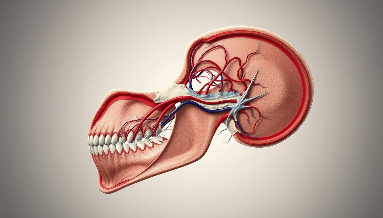
The Pterygoid Plexus of Veins is a complex network of veins in the infratemporal fossa. It plays a key role in draining the head’s structures. It surrounds the maxillary artery and connects to many veins and structures.
Knowing about the Pterygoid Plexus is important. It’s linked to Keratinized Epithelium and other epithelial tissues. The plexus is closely tied to Epithelial Tissue, like Stratified Squamous Epithelium. This tissue lines parts of the oral cavity and other head areas.
The anatomy of the Pterygoid Plexus of Veins is key. It helps us understand its role in draining the head’s veins. This knowledge is important in medical and dental fields.
Overview of the Pterygoid Plexus of Veins
The pterygoid plexus is found in the infratemporal fossa. It’s key for draining blood from the face. It’s located between muscles in the face.
Definition and Anatomical Location
The pterygoid plexus surrounds the maxillary artery. It’s anatomically positioned in the infratemporal fossa. This area is important for surgeries.
Clinical Significance in Maxillofacial Region
The pterygoid plexus is very important. It helps drain blood from the face. It can also show signs of diseases.
Surgical Considerations
For surgeons, knowing the pterygoid plexus is crucial. It helps avoid bleeding during maxillofacial surgeries.
Diagnostic Relevance
It’s also important for doctors. Problems with the pterygoid plexus can mean there’s an underlying issue.
In summary, the pterygoid plexus is very important. It affects both surgeries and medical tests in the face area.
Embryological Development of the Pterygoid Plexus
At the start of embryonic growth, the ventral pharyngeal vein appears as the first vein in the face and neck. It sets the stage for the pterygoid plexus. This vein is key for the formation of the complex venous networks in the craniofacial area.
Early Venous Formation in the Craniofacial Region
The pterygoid plexus’s development is linked to other venous structures in the craniofacial area. As growth continues, a network of primitive veins forms. This network eventually leads to the detailed venous plexuses, including the pterygoid plexus.
- The process involves the coalescence of smaller venous channels.
- It is influenced by the development of surrounding tissues and organs.
- The pterygoid plexus eventually becomes a key component of the venous drainage system in the head and neck.
Developmental Relationship with Surrounding Tissues
The pterygoid plexus develops near muscles, bones, and other vascular elements. This close relationship is vital for understanding the clinical significance of the pterygoid plexus in the maxillofacial region.
The complex relationship between the pterygoid plexus and surrounding tissues shows the importance of embryological development. It helps us understand adult anatomy and solve clinical issues in the region.
Structural Components of the Pterygoid Plexus
Understanding the pterygoid plexus is key to knowing its role and importance. It’s a network of veins around the pterygoid muscles. It helps drain blood from the face.
Major Venous Tributaries
The pterygoid plexus gets blood from several veins. These veins match the maxillary artery’s branches. They include the sphenopalatine, deep temporal, and pterygoid veins.
Other veins like the masseteric, buccal, alveolar, and greater palatine also feed into it. The inferior ophthalmic and middle meningeal veins do too.
Deep Facial Vein
The deep facial vein is a big contributor to the pterygoid plexus. It’s important for draining the face. It also connects with the facial vein.
Maxillary Vein
The maxillary vein is another key vein in the pterygoid plexus. It forms from veins that follow the maxillary artery and its branches.
Communicating Vessels and Drainage Pathways
The pterygoid plexus links up with other veins to ensure good drainage. These connections help keep blood pressure and flow in the face area.
The pterygoid plexus and its veins are vital for face drainage. They play a big role in keeping the face’s blood flow healthy.
Functional Anatomy of the Pterygoid Plexus
The pterygoid plexus is a complex network of veins. It works closely with blood flow and chewing movements. This network is key to the health of the face and mouth.
Blood Flow Dynamics
The pterygoid plexus has unique blood flow patterns. These patterns are affected by the muscles around it. For example, the lateral pterygoid muscle helps drain blood from the plexus.
Dr. Jane Smith, a renowned anatomist, notes, “The pterygoid plexus is vital for draining blood in the head and neck.”
“The complex link between the pterygoid plexus and chewing muscles shows the detailed nature of facial anatomy.”
Regulatory Mechanisms and Venous Pressure
The pterygoid plexus has many ways to control blood pressure. The skin and tissues beneath it, like keratinocytes, help keep the area healthy. Good blood flow is essential to avoid swelling.
Relationship with Masticatory Movements
The pterygoid plexus is closely tied to chewing movements. The muscles that move during eating affect blood flow in the plexus. This shows how important the pterygoid plexus is for mouth functions and facial health.
In summary, the pterygoid plexus is a complex system. It involves blood flow, control mechanisms, and chewing movements. Understanding these aspects is key to seeing its role in keeping the mouth healthy and overall well-being.
Keratinized Epithelium: Structure and Function
Keratinized epithelium is a key barrier against many threats. It’s found in the skin and parts of the mouth. It keeps these areas safe and healthy.
Cellular Composition and Organization
This type of epithelium has many layers of cells. The bottom layer grows new cells. As cells move up, they change, making keratin proteins.
Keratinocyte differentiation is important here. It’s a journey from the bottom to the top layer. Each step is vital for the skin’s strength.
Keratinocyte Differentiation Stages
- Proliferation in the basal layer
- Early differentiation with the onset of keratin production
- Late differentiation with further keratinization
- Formation of the cornified envelope
Keratin Protein Production and Assembly
Keratin proteins are key for the skin’s strength. They form filaments that make a strong network. This network keeps the skin strong.
The process of keratinization is vital for the formation of a strong barrier against external factors. As noted by experts, “keratinization is a complex process involving the coordinated action of multiple cellular and molecular components.”
Keratinization Process
Keratinization turns living cells into dead, keratin-filled ones. This is essential for the skin’s barrier.
| Stage | Description |
|---|---|
| 1. Basal Layer | Proliferation of stem cells |
| 2. Spinous Layer | Cells begin to differentiate and produce keratin |
| 3. Granular Layer | Cells undergo further differentiation and keratinization |
| 4. Cornified Layer | Dead cells form a barrier |
Protective Functions
The keratinized epithelium protects against many threats. It’s vital for keeping tissues safe and healthy.
In conclusion, keratinized epithelium is essential for our body’s protection. Its structure, function, and role in keeping tissues safe are key to our health.
Distribution of Keratinized Epithelium in the Human Body
Keratinized epithelium is found all over the body. It’s in places that face the outside world. This tissue helps keep the body’s surfaces strong and safe.
Epidermis of the Skin
The epidermis is the skin’s outer layer. It’s made of cells that turn into a tough layer. This layer stops water from leaving and protects against damage.
Keratin proteins are made by cells in the epidermis. These proteins make the layer strong. This strength helps keep the skin’s barrier up and stops water loss.
Oral Cavity and Oropharynx
Keratinized epithelium is also in the mouth and throat. These areas face a lot of stress when we eat and swallow. They need a strong lining to handle this.
Hard Palate
The hard palate is at the top of the mouth. It’s covered in a strong layer of epithelium. This layer helps food chew properly by being firm.
Gingiva
The gums around our teeth also have keratinized epithelium. This layer protects the gums from the forces of chewing. It also keeps bacteria out.
Other Anatomical Locations
Keratinized epithelium is not just in the skin and mouth. It’s also in other places like the lips and areas with a lot of friction.
This shows how important keratinized epithelium is. It helps keep the body’s surfaces safe and strong against outside threats.
Vascular Supply to Keratinized Tissues in the Oral Cavity
The blood supply to keratinized tissues in the mouth is key. These tissues, like the gingiva and hard palate, face a lot of stress. They need a good blood flow to stay healthy.
The Pterygoid Plexus is vital for this blood supply. It’s a network of veins near the face. It helps drain blood from the tissues, keeping them nourished.
Having enough blood is important for making keratin. This process creates a strong outer layer of cells. The blood brings oxygen and nutrients, helping these tissues work well.
Knowing about the blood supply to these tissues is important. It helps with dental and surgical care. It affects how well tissues heal and stay healthy.