Osteonecrosis of the Jaw (ONJ) is a serious condition. It causes bone death in the jaw. This can lead to a lot of discomfort and other problems.
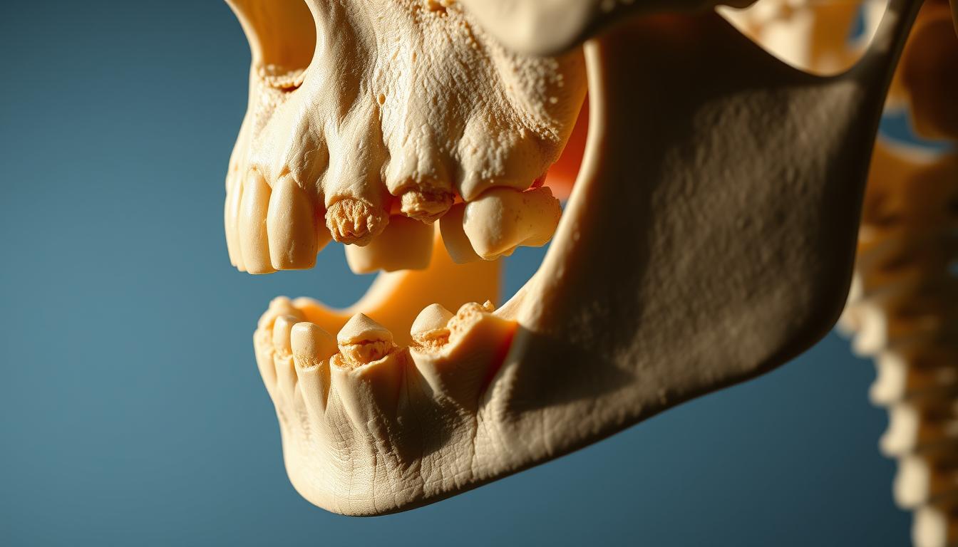
Images show how ONJ gets worse over time. They are key for doctors to diagnose and see how bad it is.
Doctors look at these images to figure out how far ONJ has spread. They then plan the best treatment. This article will cover ONJ’s causes, symptoms, and how to treat it.
Understanding Osteonecrosis of the Jaw (ONJ)
Osteonecrosis of the Jaw (ONJ) is a serious condition that affects the jawbone. It leads to significant pain and problems for those who have it.
Definition and Pathophysiology
Osteonecrosis of the Jaw is when the jawbone dies because it doesn’t get enough blood. This causes the bone to be exposed and not heal. It can happen because of certain medicines, radiation, or other health issues.
The bone’s normal repair process is disrupted. This leads to dead bone that can get infected and cause more harm.
Prevalence and Demographics
ONJ is more common in some groups than others. People taking certain medicines for osteoporosis or cancer are at higher risk. Older adults are also more likely to get it because they often have osteoporosis or cancer.
Knowing who is at risk helps us take steps to prevent ONJ.
Causes and Risk Factors of Osteonecrosis
ONJ can be caused by several factors, including medications, radiation, and health conditions. Knowing these causes is key to preventing and managing ONJ.
Medication-Related ONJ
Some medications, like bisphosphonates for osteoporosis and cancer, increase ONJ risk. Bisphosphonate-related ONJ is well-known, with higher risks in cancer patients on high doses. Denosumab, used for osteoporosis and cancer, also links to ONJ.
- High-dose bisphosphonates
- Denosumab
- Anti-angiogenic drugs
Radiation-Induced ONJ
Radiation to the head and neck raises ONJ risk. It damages bones, leading to necrosis. High doses of radiation, often for head and neck cancers, increase this risk.
Other Contributing Factors
Poor oral health, dental procedures, and conditions like diabetes can also cause ONJ. Smoking and alcohol consumption are additional risk factors.
- Poor oral hygiene
- Invasive dental procedures
- Comorbid conditions (e.g., diabetes)
Clinical Presentation of ONJ
Understanding ONJ’s clinical presentation is key for early diagnosis and treatment. The condition shows in different ways. It’s important for doctors to know its many signs and symptoms.
Early Signs and Symptoms
In the early stages, ONJ might show subtle signs that are easy to miss. These include non-specific pain, swelling, or exposed bone without infection. “Early detection is key to preventing ONJ’s progression,” studies say.
Patients might also feel joint degeneration in the affected area. This can cause discomfort and make it hard to move the jaw.
Advanced Manifestations
As ONJ gets worse, symptoms become more severe and disabling. Advanced symptoms include severe pain, significant bone exposure, and infection. This can lead to osteomyelitis or other serious issues.
The condition can greatly affect a patient’s life. It can make eating, speaking, and daily activities hard. In severe cases, surgery might be needed to manage it well.
It’s important to recognize ONJ at all stages for timely treatment and better patient outcomes.
Diagnostic Imaging Techniques for ONJ
Diagnosing Osteonecrosis of the Jaw (ONJ) needs advanced imaging. These tools help doctors see how much bone is affected. They guide treatment choices.
Panoramic Radiography
Panoramic radiography is often the first step. It shows a wide view of the jaw area. It helps spot big bone changes and dental issues.
But, it can’t see small bone changes or soft tissue problems well. So, more tests are often needed.
Computed Tomography (CT)
Computed Tomography gives detailed jaw images. It shows bone damage and sclerosis in ONJ. CT scans are great for seeing how much bone is affected.
Magnetic Resonance Imaging (MRI)
Magnetic Resonance Imaging is good at showing bone marrow and soft tissue changes. It’s great for catching early ONJ signs and soft tissue involvement. MRI helps tell ONJ apart from other jaw problems.
Cone Beam CT (CBCT)
Cone Beam CT makes detailed 3D jaw images. It shows bone structure and ONJ lesions clearly. CBCT is useful for checking ONJ’s impact on nearby structures.
Using these imaging methods, doctors can fully check ONJ. They can plan treatments and watch how the disease changes or reacts to treatment.
Radiographic Features of Early-Stage ONJ
The signs of early-stage ONJ are subtle but very important for early detection. Knowing these signs helps doctors diagnose ONJ early. This makes it easier to manage the condition.
Subtle Bone Changes
Early-stage ONJ shows small bone changes that might not be easy to see on X-rays. These changes can include osteochondral necrosis, where both bone and cartilage die. The affected area might look denser or show a slight loss of bone structure.
Doctors need to watch for these early signs closely. They can use advanced imaging to spot these small changes.
Distinguishing from Other Conditions
It’s important to tell ONJ apart from other jaw problems. Conditions like osteomyelitis and osteoradionecrosis can look similar to ONJ. This makes it hard to tell them apart.
| Condition | Radiographic Features |
|---|---|
| ONJ | Subtle bone changes, osteochondral necrosis |
| Osteomyelitis | Bone destruction, sequestra formation |
| Osteoradionecrosis | Bone necrosis, fracture, and fragmentation |
Understanding the patient’s medical history is key. Advanced imaging also helps doctors tell ONJ apart from other conditions.
Visual Characteristics of ONJ Across Different Stages
It’s important to know how ONJ looks at each stage to treat it well. ONJ changes a lot as it progresses, showing different signs at each stage.
Stage 0: At-Risk and Pre-Clinical Findings
At Stage 0, people might get ONJ because of certain treatments or radiation. Pre-clinical findings are not always easy to see. But, special imaging can spot them early.
Stage 1: Exposed Bone Without Infection
Stage 1 shows exposed bone but no infection. This is a key time to act fast to stop things from getting worse.
Stage 2: Exposed Bone With Infection
Stage 2 has exposed bone and signs of infection like pain or swelling. Osteonecrosis treatment here means fighting the infection and fixing the bone.
Stage 3: Advanced Lesions and Complications
Stage 3 has serious bone damage, maybe with fractures or fistulas. Treating Osteonecrosis here needs a big plan, possibly surgery.
| Stage | Characteristics | Treatment Approach |
|---|---|---|
| Stage 0 | At-risk, pre-clinical | Monitoring, preventive measures |
| Stage 1 | Exposed bone, no infection | Conservative management |
| Stage 2 | Exposed bone with infection | Infection management, possible surgery |
| Stage 3 | Advanced lesions, complications | Comprehensive treatment, likely surgery |
Knowing how ONJ looks at each stage is vital for the right treatment. It helps doctors manage the condition better.
Patterns of ONJ Enlargement and Progression
It’s key to know how ONJ grows and changes to manage it well. The growth is linked to dead bone, the tissue around it, and the patient’s health.
Common Expansion Pathways
ONJ lesions can grow in different ways. Necrotic bone can expose more bone, causing more damage. This often happens in the mandibular or maxillary bone, with the mandibular being more common.
| Expansion Pathway | Description | Clinical Implication |
|---|---|---|
| Direct Extension | Necrosis spreads directly to adjacent bone | Increased risk of fracture |
| Pathological Fracture | Fracture through weakened bone | Significant morbidity and possible infection |
Factors Affecting Rate of Progression
Many things can change how fast ONJ grows. These include medication-related factors like bisphosphonates, radiation therapy, and personal factors like diabetes and smoking status.
“The presence of comorbidities and the use of certain medications can significantly impact the progression of ONJ, highlighting the need for a complete treatment plan.”
A study on avascular necrosis, linked to ONJ, found that some risk factors speed up the disease. It’s important to manage these factors to slow ONJ growth.
Healthcare providers can create better plans to manage ONJ by understanding these patterns and factors. This could lead to better results for patients.
Mandibular vs. Maxillary ONJ Presentation
ONJ shows different signs in the mandible and maxilla. This is because of the bone’s structure and blood flow. These differences change how the condition looks and how it’s treated.
Anatomical Differences in Appearance
ONJ in the mandible is often worse because of its dense bone and poor blood flow. This makes it more prone to ischemic bone necrosis. On the other hand, the maxilla has more blood and thinner bone, leading to different early signs.
The way ONJ looks also varies. Mandibular lesions are usually more focused and aggressive. Maxillary lesions spread faster because of the bone’s thinness and the sinuses’ closeness.
Progression Patterns Specific to Location
ONJ grows differently in the mandible and maxilla. Mandibular ONJ gets worse faster because of its bone density and stress. Maxillary ONJ may start slower but can cause more serious problems near vital areas like sinuses and orbits.
| Characteristics | Mandibular ONJ | Maxillary ONJ |
|---|---|---|
| Initial Presentation | Often more severe, with exposed bone | May be less symptomatic initially |
| Progression Rate | Tends to progress more rapidly | Can be slower but with complex complications |
| Bone Involvement | Dense bone, more localized | Thinner bone, possible spread |
Knowing these differences helps doctors create better treatment plans. This approach improves patient outcomes by addressing the unique challenges of each location.
Differential Diagnosis Through Imaging
Imaging is key in diagnosing ONJ. It helps doctors spot the condition right. This is important for planning treatment and caring for patients.
Distinguishing ONJ from Osteomyelitis
Osteomyelitis is when bone gets inflamed due to infection. Imaging can tell ONJ apart from osteomyelitis. ONJ doesn’t show sequestra or periosteal reaction in early stages. Advanced imaging techniques like CT and MRI can spot these signs.
Differentiating from Osteoradionecrosis
Osteoradionecrosis (ORN) is a side effect of radiation therapy. It can look like ONJ on scans. But, ORN happens in areas that got radiation and shows radiation-induced damage to bone. Imaging can show cortical destruction and soft tissue swelling in ORN, not in ONJ.
Other Mimicking Conditions
ONJ can look like other conditions on scans, like metastatic disease, bone infarction, and joint degeneration. Bone infarction might look similar but lacks mucosal breakdown seen in ONJ. Careful evaluation of scans and knowing the patient’s history is key. Conditions like joint degeneration might look similar but the patient’s story helps tell them apart.
Bisphosphonate-Related ONJ: Specific Imaging Features
Imaging is key in diagnosing and managing bisphosphonate-related ONJ. This condition has unique signs that show up on X-rays. Bisphosphonates, used for osteoporosis and bone metastases, raise the risk of ONJ.
Characteristic Radiographic Findings
The signs of bisphosphonate-related ONJ can differ. They often include osteosclerosis, osteolysis, and a mix of both. These changes can be seen with panoramic radiography, CT, and MRI.
Spotting these signs early is vital for managing bisphosphonate-related ONJ well. Look for increased bone density, sequestra formation, and new bone on the periosteum.
Progression Timeline and Patterns
The growth of bisphosphonate-related ONJ can be hard to predict. Some cases stay the same for a long time, while others get worse fast. The length of bisphosphonate use, dental infections or trauma, and the patient’s health all play a role.
Knowing the progression timeline and patterns helps doctors plan better treatments. Regular imaging checks are key to spotting changes and adjusting treatment plans.
Treatment Approaches Based on Imaging Findings
Imaging is key in deciding how to treat Osteonecrosis of the Jaw (ONJ). The findings from imaging studies help doctors choose the best treatment. This depends on how severe and what the ONJ looks like.
Conservative Management
For early or mild ONJ, doctors often start with conservative management. This means using antibiotic therapy, managing pain, and watching the disease with imaging. It’s also important to teach patients about good oral hygiene and avoiding smoking.
Surgical Interventions
When early treatments don’t work, surgery might be needed. Surgeries can range from simple debridement to more complex resection of jaw parts. Imaging helps plan the surgery and check for possible complications.
Emerging Therapies
There’s ongoing research into new treatments for ONJ. Hyperbaric oxygen therapy and teriparatide show promise. These treatments aim to help healing and prevent more damage. Imaging will keep being important in checking how well these treatments work.
Monitoring ONJ Through Serial Imaging
Monitoring ONJ through serial imaging is key to understanding the disease and planning treatment. Regular imaging helps spot changes early. This allows for quick adjustments to the treatment plan.
Frequency Recommendations
The imaging frequency depends on the ONJ stage and severity. For those at high risk or with advanced disease, imaging every 3-6 months is recommended. This helps track disease progression and treatment success.
- For early-stage ONJ, imaging every 6-12 months is suggested.
- Patients on high-risk medications need regular monitoring.
Evaluating Treatment Response
Serial imaging is also vital for checking treatment response. It looks for changes in the necrotic area, bone healing, or new lesions. These signs show if the treatment is working.
Healthcare providers can then tweak treatment plans based on these changes. This ensures the best outcomes for patients.
Conclusion
Understanding Osteonecrosis of the Jaw (ONJ) is key to managing it well. Doctors use imaging to spot ONJ, track its growth, and plan treatments.
Treating ONJ requires a team effort. This includes non-surgical methods, surgery, and new treatments. Catching ONJ early is vital to stop it from getting worse and to help patients.
By knowing how ONJ looks on scans, doctors can give better care. This improves life for those with ONJ. New imaging and treatments will keep making care better.