The calcarine sulcus is a key part of the occipital lobe. It plays a big role in handling visual information. It’s linked to the visual cortex, where the primary visual cortex, or Brodmann area 17, is found.
This sulcus is key for visual perception. It helps the brain make sense of what we see. The connection between the calcarine sulcus and other parts, like the bulb of vestibule, shows how complex brain processing is.
Getting to know the calcarine sulcus function is important. It helps us understand how our brains process visual info. This makes it a big topic in neuroscience.
The Anatomy of the Calcarine Sulcus
To understand the calcarine sulcus, we must look at its anatomy. This includes where it is and the neural pathways around it. It’s a deep groove on the medial surface of the occipital lobe. This area is key for processing visual information.
Location and Structure
The calcarine sulcus starts near the top of the occipital lobe. It then runs forward until it meets the splenium of the corpus callosum. This spot is vital for its role in the visual pathway.
Kenhub explains, “the calcarine sulcus is on the medial surface of the occipital lobe.” This spot is important for processing visual data.
The calcarine sulcus has a complex structure. It’s a deep groove that separates the cuneus and lingual gyrus. These areas are key for visual processing. The sulcus is also surrounded by the primary visual cortex.
This cortex is responsible for basic visual information. It handles things like line orientation and spatial location.
Surrounding Neural Pathways
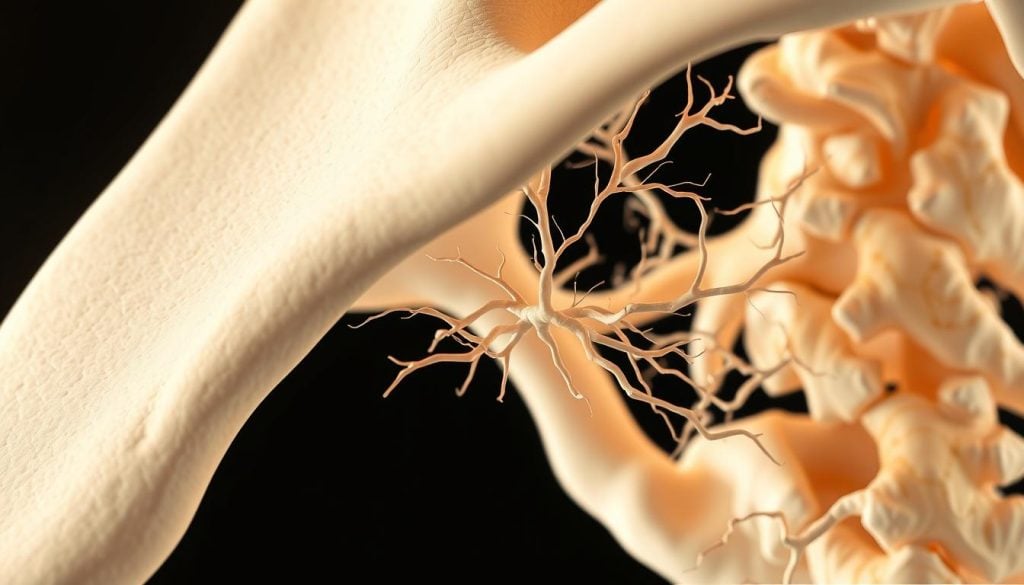
The calcarine sulcus is surrounded by important neural pathways. The primary visual cortex, around the sulcus, gets input from the lateral geniculate nucleus. It processes this information to help us understand the visual world.
| Neural Pathway | Function |
|---|---|
| Lateral Geniculate Nucleus | Transmits visual information from the retina to the primary visual cortex |
| Primary Visual Cortex | Processes basic visual information such as line orientation and spatial location |
Research shows the calcarine sulcus is key to the visual system. The neural pathways around it are vital for transmitting and processing visual information.
“The calcarine sulcus is a critical structure in the occipital lobe, playing a vital role in processing visual information.”
In conclusion, the anatomy of the calcarine sulcus is vital for understanding its role in visual processing. Its complex structure and location highlight its importance in the visual pathway.
Understanding the Bulb of Vestibule in Brain Anatomy
The bulb of vestibule is key in brain anatomy. It’s a focus in neurological studies.
Definition and Composition
The bulb of vestibule is a part of the brain. It’s important for many brain functions. It’s made of different neural tissues.
Key components of the bulb of vestibule include:
- Specific neural structures that facilitate its role in brain anatomy
- A complex network of neural pathways
- Unique cellular compositions that distinguish it from other brain regions
Evolutionary Development
The bulb of vestibule’s evolution is interesting. Studies show it evolved for important functions.
The evolutionary aspects of the bulb of vestibule can be summarized as follows:
- It has developed to support advanced neurological functions
- Its structure has adapted to the needs of the organism over time
- Comparative studies across species have provided insights into its development
Learning about the bulb of vestibule helps us understand its role in the brain. It shows its importance and function.
The Visual Processing Role of the Calcarine Sulcus
The calcarine sulcus is key for understanding visual data. It contains the primary visual cortex, also known as Brodmann area 17. This area is in charge of handling visual information.
Primary Visual Cortex Integration
The primary visual cortex, found in the calcarine sulcus, is the first stop for visual data in the brain. It gets input from the lateral geniculate nucleus of the thalamus. This nucleus sends visual info from the retina.
This integration is vital for the first steps in processing visual stimuli. It includes line orientation, spatial location, and movement.
The primary visual cortex is set up in a way that mirrors the retina. This means the neurons are arranged like the photoreceptors in the retina. This setup is key for keeping visual information in the right order as it’s processed.
Signal Processing Mechanisms
Signal processing in the calcarine sulcus is complex. The primary visual cortex has different types of cells. Simple cells focus on specific orientations and spatial frequencies. Complex cells, on the other hand, handle more complex patterns, like movement in a certain direction.
Signal processing mechanisms in the calcarine sulcus do more than just detect simple features. The primary visual cortex also handles more complex tasks. It integrates information from different parts of the visual field and processes visual stimuli in context.
How the Bulb of Vestibule Contributes to Visual Perception
The bulb of vestibule is not directly linked to seeing things. Yet, it plays a role in brain anatomy. Knowing its part can help us understand how we see the world.
Neural Connectivity Patterns
The bulb of vestibule is part of the vestibular system. This system helps us balance and know where we are in space. It connects with other brain areas, which might affect how we see things.
Research is looking into how the vestibular system links to the visual cortex. It’s found that balance and spatial awareness can influence how we process visual information.
Information Transmission Processes
Signals from the bulb of vestibule travel to other brain parts through complex pathways. These paths help mix vestibular info with what we see. This mix helps us understand our surroundings better.
| Structure | Function | Relation to Visual Perception |
|---|---|---|
| Bulb of Vestibule | Part of the vestibular system, involved in balance and spatial orientation | Indirectly influences visual perception through neural connectivity |
| Vestibular System | Processes information related to balance and movement | Affects visual processing, specially in spatial awareness tasks |
| Visual Cortex | Primary region for processing visual information | Integrates vestibular inputs to enhance visual perception |
Learning about the bulb of vestibule’s role in seeing things shows how brain parts work together. More research could uncover more about how we see the world.
The Relationship Between the Calcarine Sulcus and Visual Field Mapping
The connection between the calcarine sulcus and visual field mapping is very interesting. The calcarine sulcus is a deep groove in the back of the brain. It’s key for handling visual information.
Retinotopic Organization
The calcarine sulcus is part of retinotopic organization. This means the visual field is mapped onto the brain in a way that matches the retina. This mapping helps the brain understand visual signals well.
Research shows the calcarine sulcus has a precise map of the visual field. This is important for recognizing objects and seeing how things are arranged in space.
Visual Field Representation
The calcarine sulcus is also important for visual field representation. The top and bottom parts of the sulcus deal with different parts of what we see. The top part handles the lower part of our visual field, and the bottom part handles the upper part.
- The calcarine sulcus processes visual information from the opposite side of our visual field.
- This structure is vital for combining visual information from all parts of our field of vision.
- Damage to the calcarine sulcus can cause specific problems with seeing.
In summary, the calcarine sulcus is essential for mapping the visual field. It does this through its role in retinotopic organization and visual field representation. Understanding this is key to learning about how we see and the brain’s role in it.
Functional Neuroimaging Studies of the Calcarine Sulcus
Studies using functional neuroimaging have given us a better understanding of the calcarine sulcus. It’s a key part of the primary visual cortex. Different imaging techniques have helped us learn how it works.
fMRI Findings
Functional MRI (fMRI) has been key in figuring out the calcarine sulcus’s role. It shows the sulcus is active when we do visual tasks. fMRI findings have consistently shown a high degree of retinotopic organization within the calcarine sulcus, showing how visual inputs are mapped.
With fMRI, researchers can see how the visual field is mapped onto the calcarine sulcus. This mapping is vital for understanding how the brain processes visual information.
PET Scan Insights
PET scans have given us more insights into the calcarine sulcus’s activity. PET scan studies have shown increased blood flow and glucose metabolism in the calcarine sulcus during visual tasks. This shows the sulcus is very active when we’re processing visual information.
These studies, including fMRI and PET scans, have greatly improved our knowledge of the calcarine sulcus. The detailed insights from these studies are important for both research and clinical use.
Clinical Significance of the Bulb of Vestibule
It’s key to know how the bulb of vestibule affects health. This part of the vestibular system plays a big role in many health issues. It’s important for diagnosing and treating problems.
Associated Pathologies
The bulb of vestibule is linked to several health issues. These include:
- Vestibular neuritis, which causes inflammation and problems with the vestibule.
- Meniere’s disease, marked by vertigo, tinnitus, and hearing loss.
- Benign paroxysmal positional vertigo (BPPV), causing brief but intense vertigo.
These conditions show how vital the bulb of vestibule is for our balance. Damage here can lead to serious balance problems.
Diagnostic Approaches
Diagnosing issues with the bulb of vestibule involves several steps. These include:
- Vestibular function tests, like electronystagmography (ENG) and videonystagmography (VNG).
- Imaging studies, such as MRI and CT scans, to see the vestibule and related areas.
- Tests to check balance and equilibrium to see how well the vestibule is working.
Recent studies highlight the importance of understanding the bulb of vestibule. This knowledge is vital for accurate diagnosis and treatment of vestibular disorders. A StatPearls publication notes, “Understanding the clinical significance of the bulb of vestibule is essential for diagnosing and treating related pathologies.”
The bulb of vestibule’s role is complex. It’s involved in both normal balance and health issues. More research will help us better manage vestibular disorders.
Lesions and Deficits: When the Calcarine Sulcus is Damaged
Damage to the calcarine sulcus can lead to serious visual problems. This area is key in our visual pathway. Lesions here can cause big visual issues.
Visual Field Defects
Damage to the calcarine sulcus can cause visual field defects. This means some parts of what we see are affected. It’s because this area helps process what the retina sees.
Lesions here can lead to hemianopia, where half of what we see is lost. The exact defect depends on where and how much damage there is. Knowing this helps doctors find the problem and treat it.
Cortical Blindness
In severe cases, damage can cause cortical blindness. This is when we lose all visual perception because of damage to both sides of the primary visual cortex. It’s a rare condition that can happen from stroke, trauma, or infection.
People with cortical blindness might keep their pupillary reflexes. This is because the damage is in the visual cortex, not the retina or optic nerve. Treatment aims to help them use what little vision they have left.
Developmental Aspects of the Calcarine Sulcus
The calcarine sulcus is key for seeing things. It grows a lot before and after birth. This growth helps us understand how it works with visual information.
Prenatal Formation
The calcarine sulcus starts to form in the womb. It happens when the brain’s surface folds. Research says it begins around 24 weeks of pregnancy.
Postnatal Maturation
After birth, the calcarine sulcus keeps growing. This growth is important for seeing clearly. It happens in the first years of life.
Genes and what we see play big roles in this growth. Knowing how the calcarine sulcus develops helps us understand vision better. It shows how important early experiences are for our vision.
Comparative Anatomy: The Calcarine Sulcus Across Species
Looking at the calcarine sulcus in different species shows its key role in evolution. This sulcus is vital for how we see things. Its presence in many species shows it’s very important.
Primate Comparisons
Research has found that primates have a calcarine sulcus. This shows that this part of the brain is the same in these species. It means they share a way of processing visual information that has stayed the same over time.
Evolutionary Significance
The calcarine sulcus is key for how we see. Its presence in many species shows it’s essential for understanding what we see.
| Species | Presence of Calcarine Sulcus | Visual Processing Characteristics |
|---|---|---|
| Humans | Present | Complex visual processing, including object recognition |
| Primates | Present | Similar to humans, with advanced visual capabilities |
| Other Mammals | Varies | Different levels of visual processing complexity |
Studying the calcarine sulcus in different species shows it’s been important for a long time. It helps us understand how our brains process what we see.
Advanced Imaging Techniques for Studying the Bulb of Vestibule
Advanced imaging has changed how we study the bulb of vestibule. It gives us new insights into its structure and role. The bulb of vestibule is key for balance and keeping our body steady.
Studies now use new imaging methods to learn more about the bulb of vestibule. Techniques like diffusion tensor imaging (DTI) and functional connectivity analysis are very helpful.
Diffusion Tensor Imaging
DTI is a way to see brain connections with MRI. It helps researchers understand how the bulb of vestibule connects to other parts of the balance system. This is important for knowing how it helps us stay balanced.
Functional Connectivity Analysis
Functional connectivity analysis looks at how brain areas talk to each other. It shows how the bulb of vestibule works with other brain parts. This helps scientists understand how it helps with balance.
Using DTI and functional connectivity together gives us a full picture of the bulb of vestibule. These methods help doctors diagnose and treat balance problems better.
The Role of the Calcarine Sulcus in Higher Visual Processing
The calcarine sulcus is key for advanced visual tasks like recognizing objects and being aware of what we see. It’s found in the occipital lobe and is vital for making sense of complex visual information.
Object Recognition Contributions
The calcarine sulcus helps a lot with recognizing objects. It takes visual info from the retina and sends it to higher areas for processing. This includes the primary visual cortex, where it plays a big part in understanding visual signals.
Object recognition is complex and needs many visual features. The calcarine sulcus is essential for combining these features.
Visual Awareness Mechanisms
The calcarine sulcus also affects how we become aware of visual information. It helps us see and understand what’s around us. Studies show it’s active when we’re consciously aware of visual stimuli.
In summary, the calcarine sulcus is vital for advanced visual tasks like recognizing objects and being aware of our surroundings. Knowing its role helps us understand how we perceive and process visual information.
Future Research Directions in Calcarine Sulcus Function
Our understanding of the calcarine sulcus is growing, thanks to new technologies. This area is key for how we see the world. It’s a big focus in neuroscience.
Emerging Technologies
New tools like functional MRI (fMRI) and diffusion tensor imaging (DTI) are helping us learn more. These emerging technologies are essential for studying the brain’s visual networks.
Unanswered Questions
Even with all we’ve learned, there’s more to discover. For example, how does the calcarine sulcus work with other brain areas? Like the vestibule bulb, to make sense of what we see?
| Research Area | Current Status | Future Directions |
|---|---|---|
| Neuroimaging Techniques | Advances in fMRI and DTI | Improved resolution and analysis methods |
| Neural Connectivity | Understanding calcarine sulcus interactions | Investigating connectivity with vestibule bulb |
Looking ahead, research will blend new tech with a dive into the unknown. This will help us understand the calcarine sulcus better.
Conclusion: The Essential Role of the Calcarine Sulcus and Bulb of Vestibule in Visual Processing
The Calcarine Sulcus is key to how we see the world. It’s a vital part of the brain’s visual center. Working with the Bulb of Vestibule, it helps us understand and process what we see.
Learning about the Calcarine Sulcus helps us understand how we see. Studies show it plays a big role in how our brain handles visual information. The term Light Bulb might not directly relate to brain functions, but it shows the excitement of discovering how our brain works.
The connection between the Calcarine Sulcus and how we map our visual field is important. Knowing how these parts work helps scientists and doctors. They can then find better ways to treat vision problems.