Noonan syndrome (NS) is a genetic disorder. It has unique facial features, heart defects, and varying levels of intellectual disability. Spotting these signs is key for correct diagnosis and care.
Spotting Noonan syndrome is vital for better patient care and treatment. Knowing the signs of NS helps doctors give the right help.
This guide looks at pictures of patients with Noonan syndrome. It shows the main signs that help doctors diagnose it. Our goal is to improve understanding and spotting of this complex genetic disorder.
The Spectrum of Noonan Syndrome
Understanding Noonan syndrome means looking at its definition, how common it is, and its history. It’s a genetic disorder that affects many body systems. It happens because of problems with the RAS-MAPK signaling pathway.
Definition and Prevalence
Noonan syndrome has many symptoms and affects about 1 in 1000 to 1 in 2500 babies. It’s known for its unique face, heart issues, and delays in development.
“Noonan syndrome is a common genetic disorder,” doctors say. “It needs a careful look for diagnosis.” This shows why doctors must be aware of it.
Historical Context and Clinical Recognition
The first signs of Noonan syndrome were seen in the 1960s by Dr. Jacqueline Noonan. She noticed a pattern of features in patients. Over time, we’ve learned more about its genetics and symptoms.
Doctors have gotten better at spotting Noonan syndrome. Getting a diagnosis early is key to managing it well.
Noonan-like Features: Clinical Overview
Spotting Noonan-like features early is key for diagnosing and treating Noonan syndrome. These signs include physical traits and developmental milestones. It’s important for doctors to know these well.
Defining Characteristics
Noonan-like traits include many facial and physical oddities. Some main signs are:
- Facial dysmorphology, such as hypertelorism and ptosis
- Short stature and growth delays
- Congenital heart defects, like pulmonary valve stenosis
- Developmental delay and learning disabilities
These traits can differ a lot from person to person. So, a detailed check-up is needed.
Variability in Expression
The way Noonan-like traits show up can vary a lot. Some people might have mild signs, while others have more obvious ones. This is because of the genetic variety in Noonan syndrome.
Age-Related Changes in Presentation
Noonan-like traits change as people get older. Facial signs are often most noticeable in young and middle childhood. As people grow, some signs may fade, while others stay or change.
Knowing these age-related changes is important for correct diagnosis and care at any age.
Genetic Basis of Noonan Syndrome
The RAS/MAPK signaling pathway is key to understanding Noonan syndrome. It controls cell growth, development, and survival. Mutations in this pathway cause Noonan syndrome.
RAS/MAPK Pathway Disorders
Noonan syndrome falls under RASopathy, a group of disorders. These disorders share features like facial abnormalities, heart issues, and developmental delays. The RAS/MAPK pathway is vital for cell functions, and its problems lead to Noonan syndrome symptoms.
Key genes involved in the RAS/MAPK pathway include PTPN11, SOS1, RAF1, and KRAS.
PTPN11 and Other Gene Mutations
PTPN11 mutations are common in Noonan syndrome, found in about 50% of cases. PTPN11 encodes the SHP2 protein, a key RAS/MAPK pathway regulator. SOS1, RAF1, and KRAS are also linked to Noonan syndrome. Finding these mutations helps in diagnosis and understanding the condition’s variability.
“The genetic heterogeneity of Noonan syndrome highlights its complex diagnosis and the need for detailed genetic testing.”
Inheritance Patterns and Genetic Counseling
Noonan syndrome is usually inherited in an autosomal dominant pattern. This means one mutated gene copy is enough to cause the condition. Genetic counseling is vital for families, providing information on risk and variability.
Genetic counseling also talks about the meaning of genetic tests. It explains the importance of ongoing care and management for Noonan syndrome.
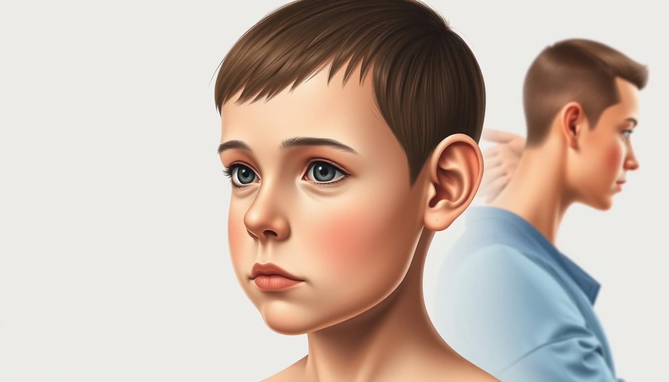
Craniofacial Dysmorphology
Craniofacial dysmorphology in Noonan Syndrome includes many distinct facial features. These are key for diagnosing and understanding the condition.
Hypertelorism and Ocular Abnormalities
Hypertelorism is a hallmark of Noonan Syndrome. It means the eyes are farther apart than usual. This can be paired with other eye issues like down-slanting palpebral fissures and strabismus. These traits make the face of someone with NS unique.
Ptosis and Epicanthal Folds
Ptosis, or a drooping eyelid, is common in NS. It often goes with epicanthal folds, which are skin folds at the eyes’ inner corners. These features greatly affect facial expression and are key signs for diagnosis.
Low-Set and Posteriorly Rotated Ears
People with Noonan Syndrome often have low-set ears that are posteriorly rotated. This ear shape is a big part of the facial dysmorphology seen in NS.
Clinical Imaging Techniques for Facial Features
Getting accurate pictures of these facial traits is vital for diagnosis and tracking. Clinical imaging techniques, like 2D and 3D photography, are used to capture these details. These images help doctors see how severe the dysmorphology is and track any changes.
Here’s a list of main points about craniofacial dysmorphology in Noonan Syndrome:
- Hypertelorism and ocular abnormalities
- Ptosis and epicanthal folds
- Low-set and posteriorly rotated ears
- Use of clinical imaging for documentation
Distinctive Facial Characteristics
Noonan Syndrome is known for its unique facial traits. These traits help doctors diagnose and keep track of the condition. The face of someone with Noonan Syndrome looks quite different from others.
Triangular Face Shape
A triangular face shape is a key sign of Noonan Syndrome. This shape is often seen with a broad forehead and a pointed chin. It makes the face look triangular.
High Forehead and Widow’s Peak
Many people with Noonan Syndrome have a high forehead. Some also have a widow’s peak hairline. These traits make their face stand out.
Micrognathia and Dental Anomalies
Micrognathia, or a small lower jaw, is common in Noonan Syndrome. It can cause dental problems and tooth alignment issues. This might need orthodontic treatment.
Photographic Documentation Methods
Photos are key for tracking facial changes in Noonan Syndrome. Using the same photography methods helps compare pictures over time.
| Feature | Description | Clinical Significance |
|---|---|---|
| Triangular Face Shape | Broader forehead, pointed chin | Aids in diagnosis |
| High Forehead | Often with widow’s peak | Contributes to distinctive appearance |
| Micrognathia | Small lower jaw | May lead to dental issues |
A clinical expert says, “Photos help track facial changes over time. This is key for managing Noonan Syndrome.”
“Using the same photo methods is important for accurately tracking facial changes over time.”
Neck and Thoracic Manifestations
Neck and thoracic signs are important in Noonan Syndrome. They help doctors diagnose and manage the condition. These signs can greatly affect a person’s life quality.
Webbed Neck (Pterygium Colli)
A webbed neck, or pterygium colli, is a common sign of Noonan Syndrome. It happens when the neck skin forms a web-like shape at birth.
- It shows as extra skin folds in the neck.
- It might make moving the neck harder.
- Surgery might be needed for looks or to improve movement.
Low Posterior Hairline
People with Noonan Syndrome often have a low posterior hairline. This is a small but clear sign. It’s a key clue for doctors to diagnose.
Pectus Excavatum and Carinatum
Pectus excavatum and pectus carinatum are common chest problems in Noonan Syndrome. These issues affect the chest shape and can harm breathing and heart work.
Radiographic evaluation is key to check chest problems in Noonan Syndrome. Tests like chest X-rays and CT scans help doctors see how bad the chest issues are. This helps them decide the best treatment.
- First, doctors usually do a chest X-ray.
- CT scans give more detailed info on the chest shape and how bad the problem is.
- What the tests show helps doctors choose if surgery or other treatments are needed.
Growth Patterns and Developmental Milestones
It’s key to know about growth patterns and milestones for Noonan Syndrome. People with NS often grow shorter and may develop slower. Keeping an eye on their growth is very important.
Short Stature and Growth Charts
Many with Noonan Syndrome grow shorter. Growth charts help track their growth. Regular checks and growth charts help spot growth problems early.
Developmental Delay Spectrum
People with NS often hit milestones like sitting and walking later. The delay can vary a lot. Each person needs their own plan and support.
Puberty and Hormonal Considerations
Puberty in NS can bring hormonal issues. Watching for puberty signs and checking hormone levels is key. Sometimes, growth hormone therapy is used to help with height.
Growth Monitoring Techniques
Tracking growth means taking regular measurements. This includes height, weight, and head size. These are plotted on charts to see how they’re doing. Sometimes, bone age tests are used too.
| Growth Monitoring Technique | Description | Frequency |
|---|---|---|
| Height Measurement | Assessing standing height | Every 6 months |
| Weight Measurement | Assessing body weight | Every 6 months |
| Bone Age Assessment | Evaluating skeletal maturity | Annually |
Cardiac Abnormalities and Imaging
Cardiac issues are a big worry for people with Noonan Syndrome. They need detailed imaging and tests to find and manage these problems. Early detection and treatment are key to improving their quality of life.
Pulmonary Valve Stenosis
Pulmonary valve stenosis is common in Noonan Syndrome. It happens when the pulmonary valve gets too narrow. This blocks blood flow from the right ventricle to the pulmonary artery. Echocardiography helps diagnose this by looking at the valve’s shape and how it works.
Hypertrophic Cardiomyopathy
Hypertrophic cardiomyopathy (HCM) is also seen in NS patients. It makes the heart muscle too thick, which can block blood flow. Cardiac MRI is vital for spotting HCM. It gives clear pictures of the heart’s structure and how it works.
Other Structural Heart Defects
NS patients can also have other heart defects like atrial septal defects and ventricular septal defects. Detailed heart imaging is needed to find these and plan treatment.
Echocardiographic and MRI Findings
Echocardiography and MRI are essential for diagnosing heart problems in Noonan Syndrome. Echocardiography shows the heart in real-time, checking valves and structure. MRI gives detailed pictures of the heart’s anatomy, helping spot complex issues and plan surgeries.
Using both echocardiography and MRI findings is key for managing heart issues in NS patients. This approach helps doctors understand the heart’s condition better. It guides treatment choices and improves patient outcomes.
Skeletal and Limb Features
People with Noonan Syndrome often have skeletal and limb issues. These signs are key for diagnosis and care. The range of skeletal problems includes various deformities and structural issues.
Cubitus Valgus
Cubitus valgus is a common elbow deformity in NS patients. It causes an abnormal elbow angle. Early identification is important for managing the condition.
Chest Wall Deformities
Chest wall issues like pectus excavatum or carinatum are common. These can affect breathing and heart function. Comprehensive evaluation is needed to decide if treatment is necessary.
Scoliosis and Spinal Issues
Scoliosis, or spine curvature, is a significant skeletal feature. Regular checks are key to catch any worsening and plan treatment.
- Scoliosis can cause serious spinal deformity if not treated.
- Early detection through X-rays is critical.
Radiographic Assessment Techniques
Radiographic assessment is essential for checking skeletal issues. X-rays and other imaging help measure deformity extent.
Hematological and Lymphatic Manifestations
Hematological and lymphatic issues are common in Noonan Syndrome. They need careful evaluation and management. These problems can greatly affect a patient’s life and health outcomes.
Bleeding Disorders
Bleeding disorders are a big worry in Noonan Syndrome. Patients might bruise easily, have nosebleeds, and face other bleeding issues. This is because of problems with blood clotting or platelet function.
Lymphatic Abnormalities
Lymphatic problems, like lymphedema and lymphatic dysplasias, are also common. These can cause swelling, pain, and a higher chance of infections.
Hematological Malignancies
People with Noonan Syndrome are more likely to get blood cancers, like juvenile myelomonocytic leukemia (JMML). It’s important to watch them closely for early signs.
Laboratory and Imaging Evaluation
Lab tests, like complete blood counts and coagulation studies, are key for diagnosing and treating blood issues. Imaging, such as ultrasound and MRI, helps check lymphatic problems.
| Manifestation | Diagnostic Approach | Management Strategy |
|---|---|---|
| Bleeding Disorders | Coagulation studies, platelet function tests | Factor replacement, platelet transfusions |
| Lymphatic Abnormalities | Ultrasound, MRI lymphography | Compression therapy, surgical intervention |
| Hematological Malignancies | Bone marrow biopsy, genetic testing | Chemotherapy, targeted therapy |
Neurological and Cognitive Assessment
Noonan Syndrome often involves a range of neurological and cognitive impairments. These need a thorough evaluation. People with NS may have varying degrees of developmental delay, intellectual disability, and learning disabilities.
Intellectual Disability Spectrum
Intellectual disability in NS can range from mild to severe. Early intervention is key to managing the condition. It helps improve the quality of life for those affected.
Learning Disabilities
Learning disabilities are common in NS, affecting academic and social development. Tailored educational programs can greatly benefit these individuals.
Motor Skills and Coordination
Motor skills and coordination can be affected, leading to difficulties in physical activities. Physical therapy is often recommended to improve motor function.
Neuroimaging Findings
Neuroimaging plays a vital role in assessing neurological features associated with NS. Techniques such as MRI can help identify structural brain abnormalities.
The insights gained from neuroimaging, combined with clinical evaluation, aid in the management of NS. They address both neurological and cognitive aspects.
Clinical Photography in Documenting Noonan-like Features
Clinical photography is key in documenting Noonan-like features. It provides a visual record that helps in diagnosis and tracking. This visual documentation is vital for clinicians to see the presence and growth of features linked to Noonan syndrome.
Standardized Photography Protocols
Standardized photography protocols are vital for consistent and useful clinical photos. They ensure the use of the same lighting, camera settings, and patient position. Standardization makes it easier to compare images over time and between patients.
A study on genetic disorders highlighted the need for a standardized method to capture facial features. These features are often key in diagnosing conditions like Noonan syndrome.
Sequential Imaging for Monitoring
Sequential imaging is key for tracking the growth of Noonan-like features over time. It lets clinicians see changes, check if treatments are working, and make better care decisions.
Regular photos can show small changes that might not be seen during regular check-ups.
| Feature | Baseline | Follow-Up |
|---|---|---|
| Facial Dysmorphology | Present | Progressive |
| Craniofacial Abnormalities | Noted | Stable |
Ethical Considerations in Clinical Photography
Ethical issues are critical in clinical photography, more so in pediatric and sensitive cases. Patient consent, privacy, and confidentiality must be respected. Following institutional guidelines and regulations is essential.
Digital Image Analysis Techniques
Digital image analysis techniques are getting better, allowing for more detailed photo assessments. Software can measure facial proportions, spot small changes, and analyze other features important for Noonan syndrome.
By using standardized protocols, sequential imaging, and advanced digital analysis, clinicians can improve diagnosis and care for patients with Noonan-like features.
Differential Diagnosis Through Imaging
Imaging is key in diagnosing Noonan Syndrome (NS). It helps tell NS apart from similar conditions like Turner syndrome and Williams syndrome.
Turner Syndrome
Turner syndrome affects females, causing short stature and delayed puberty. It also has unique physical traits. Echocardiography and ultrasound are used to spot heart problems and other signs of Turner syndrome.
Williams Syndrome
Williams syndrome is a genetic disorder with special facial features and heart issues. Echocardiography is vital for finding heart problems in Williams syndrome.
Other RASopathies
NS is part of the RASopathies group, which includes other syndromes. These syndromes share similar traits, making diagnosis tricky.
Comparative Imaging Features
Imaging can help tell these syndromes apart. For example:
- Cardiac abnormalities: NS often has pulmonary valve stenosis, while Turner syndrome might have a bicuspid aortic valve.
- Craniofacial features: Williams syndrome has an “elfin” face, while NS has a triangular face.
- Skeletal features: Turner syndrome often has cubitus valgus, while NS might have pectus excavatum or carinatum.
By looking at these imaging features, doctors can make a more precise diagnosis. This helps in creating the right treatment plan.
Advanced Imaging Modalities in Diagnosis
Advanced imaging has greatly improved how we diagnose and manage Noonan Syndrome (NS). These new technologies give us detailed views of NS’s complex symptoms. This helps doctors make more accurate diagnoses and create better treatment plans.
3D Facial Analysis
Three-dimensional facial analysis is a new way to measure facial differences in NS. It can spot small changes in facial shape. This helps doctors identify the typical signs of NS.
Cardiac MRI and CT Applications
Cardiac MRI and CT scans are key for checking heart problems in NS patients. They give clear pictures of the heart. This helps find issues like pulmonary valve stenosis and hypertrophic cardiomyopathy.
| Imaging Modality | Application in NS |
|---|---|
| Cardiac MRI | Assessment of cardiac structure and function |
| Cardiac CT | Detailed imaging of coronary arteries and cardiac anatomy |
Neuroimaging Considerations
Neuroimaging is important for looking at NS’s brain effects. MRI can spot brain problems. It helps doctors plan the best care.
Future Imaging Technologies
New imaging tech, like advanced MRI and AI, will help even more. They might give us even clearer views of NS. This could lead to better care for patients.
Diagnostic Approach and Genetic Testing
To diagnose Noonan Syndrome (NS), doctors use a mix of clinical checks and genetic tests. This method is key to correctly identifying the condition. It helps understand its effects on the patient and their family.
Clinical Diagnostic Criteria
Doctors look for specific signs to diagnose NS. These include unique facial features, heart problems, and delays in development. Clinical diagnostic criteria help figure out if someone has NS.
- Facial dysmorphology
- Congenital heart defects
- Developmental and growth delays
Genetic Testing Strategies
Genetic tests are vital to confirm NS. Mutations in several genes, like PTPN11, SOS1, and RAF1, are linked to NS. The type of test used depends on the symptoms and family history.
Prenatal Diagnosis
Prenatal diagnosis is possible if the family’s genetic history shows NS. This means testing the fetus for the known mutation.
Interpretation of Genetic Results
Understanding genetic results needs special knowledge. Genetic counselors are essential. They explain the results and their meaning to the family.
Management and Multidisciplinary Care
Noonan Syndrome is complex and needs a team approach for care. A group of healthcare experts work together to meet the needs of patients.
Growth Hormone Therapy
Growth hormone therapy is used for growth delays in Noonan Syndrome. It helps improve growth and development.
Cardiac Management
Heart problems are common in Noonan Syndrome. Cardiac care is key. This includes monitoring and treatments like surgery or medicine.
Developmental Interventions
Developmental therapies are vital for Noonan Syndrome. They include physical, occupational, and speech therapy. These help with developmental delays and improve life quality.
Long-term Monitoring Protocols
Long-term monitoring is vital for Noonan Syndrome. Regular check-ups with a team help catch and treat new issues quickly.
| Aspect of Care | Interventions | Frequency |
|---|---|---|
| Growth Monitoring | Growth hormone therapy, nutritional support | Every 6 months |
| Cardiac Care | Regular echocardiograms, medication, surgery | Annually or as needed |
| Developmental Support | Physical, occupational, speech therapy | Ongoing, adjusted based on need |
Conclusion
It’s important to spot Noonan-like features early for diagnosing and managing Noonan syndrome (NS). This genetic disorder has unique facial traits, heart issues, and developmental delays. These signs are key to understanding NS.
Knowing the genetic roots of NS is vital for counseling and testing. A team of cardiologists, geneticists, and developmental specialists is needed. They work together to tackle the many challenges of NS.
Healthcare teams should be aware of how NS can change over time. This knowledge helps in providing better care and improving life for those with NS. Ongoing research will help us better understand and manage NS.