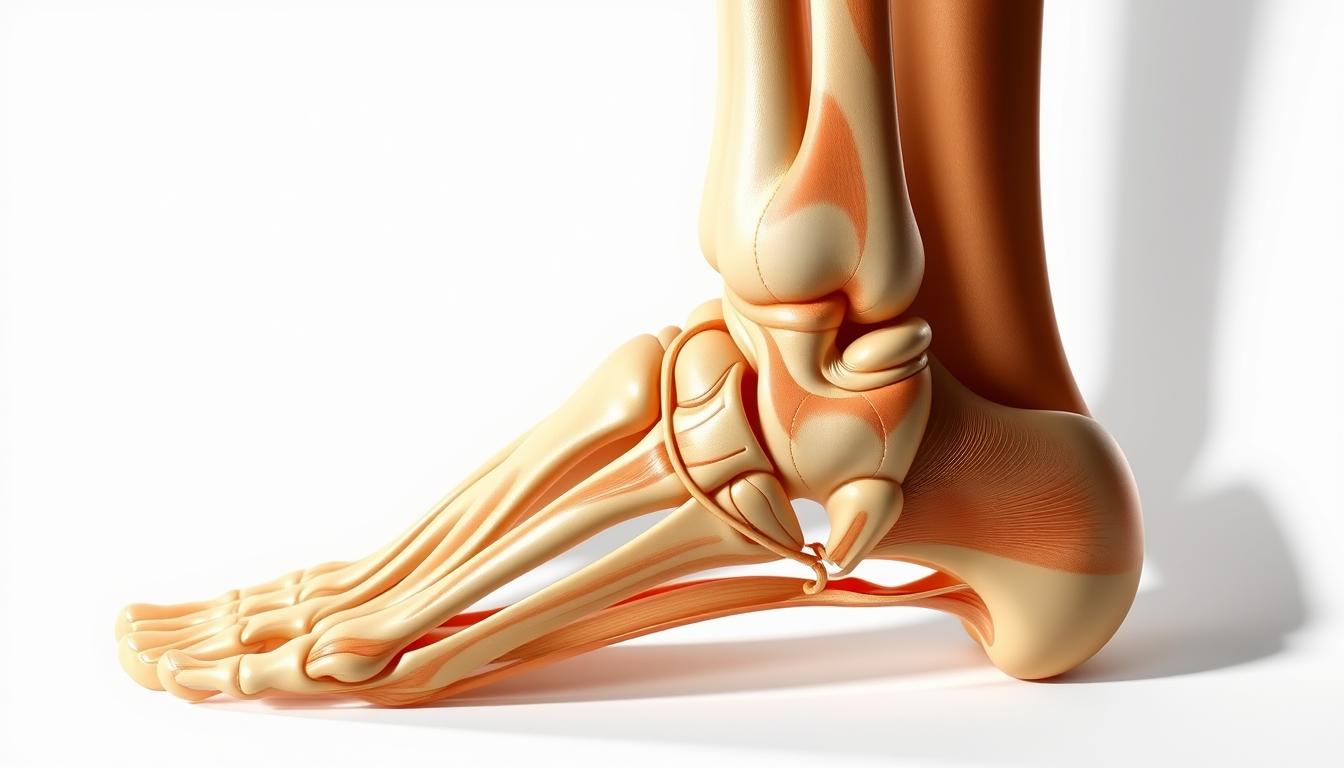
Understanding the dorsolateral view of the left foot and ankle is key for medical experts and those interested in foot anatomy. The foot’s structure is complex, with many bones, muscles, and ligaments. It’s both fascinating and challenging to study.
The ankle joint, or talocrural joint, is important for movements like dorsiflexion and plantar flexion. Knowing the foot’s anatomy well helps in diagnosing and treating foot problems.
The dorsolateral view gives a special look at the foot’s structure. It shows how all parts work together. This view helps us understand the foot’s complexity and its role in our daily movements.
The Anatomical Perspective of the Foot
The study of the foot’s anatomy gives us insights into its function and structure. Knowing how the foot is built helps doctors diagnose and treat foot problems.
Defining the Dorsolateral View
The dorsolateral view shows the foot from the top and outer side. It’s key for seeing how foot bones fit together with muscles and ligaments. This view helps us understand the foot’s complex design.
Anatomical Planes and Terminology
To talk about the foot’s anatomy, we need to know the anatomical planes and terms. The foot can be seen from different angles, like the sagittal, frontal, and transverse planes. Knowing these terms helps doctors communicate better.
Clinical Significance of Dorsolateral Perspective
In clinics, the dorsolateral view is very important for diagnosing and treating foot issues. It helps doctors check if the foot structure is right. This view is key for spotting problems like fractures, sprains, and tendon injuries.
Understanding the anatomical perspective of the foot helps doctors make accurate diagnoses and treatment plans. The dorsolateral view is a big part of this, giving a detailed look at the foot’s anatomy.
Osteology of the Ankle Complex
The study of the ankle complex focuses on the distal tibia and fibula, talus, and calcaneus. These bones are key to movement and supporting the body’s weight.
Distal Tibia and Fibula
The distal tibia and fibula connect the lower leg to the foot. The distal tibia’s medial malleolus and the fibula’s lateral malleolus are vital for ankle stability. The fibula, though smaller, is essential for the ankle’s lateral side.
Talus and Calcaneus
The talus connects the leg to the foot, enabling movement. It links with the distal tibia and fibula at the talocrural joint. The calcaneus, or heel bone, is the largest tarsal bone. It supports muscles and ligaments.
| Bone | Characteristics | Function |
|---|---|---|
| Distal Tibia | Forms medial malleolus | Provides medial stability |
| Distal Fibula | Forms lateral malleolus | Provides lateral stability |
| Talus | Articulates with tibia and fibula | Facilitates ankle movement |
| Calcaneus | Largest tarsal bone | Attachment site for muscles and ligaments |
Articulations of the Ankle
The ankle complex has several joints that allow for movement. The talocrural joint is key for dorsiflexion and plantarflexion. Knowing these joints is important for ankle health.
Tarsal Bones from the Dorsolateral Perspective
The tarsal bones are key to the foot’s movement and structure. They make up the back part of the foot. This is important for how the foot works.
Cuboid and Navicular
The cuboid bone is on the outside of the foot. It’s between the calcaneus and the fourth and fifth metatarsals. It helps form the lateral longitudinal arch.
The navicular bone is on the inside. It connects with the talus and the cuneiform bones. This helps the foot move in complex ways.
Cuneiform Bones
The cuneiform bones are in front of the navicular bone. They connect with the navicular and the metatarsals. These bones help form the arches of the foot, allowing for movement.
Tarsal Articulations and Movements
The tarsal bones have many important connections. These connections allow the foot to move in different ways. Key connections include:
- The talocalcaneal joint, which allows for inversion and eversion.
- The talonavicular joint, which is key for the foot’s mobility.
- The calcaneocuboid joint, which helps with lateral stability.
Together, these connections help the foot move in complex ways. This includes inversion, eversion, dorsiflexion, and plantarflexion.
Metatarsals and Phalanges
The metatarsals and phalanges are key parts of the foot. They help with toe movement and foot function. Together, they give the foot stability, support, and mobility.
Metatarsal Structure and Function
The metatarsals are five long bones in the foot. They link the toes to the rest of the foot. They are vital for weight-bearing and the foot’s arch.
The first metatarsal bone is the thickest and shortest. It bears a lot of body weight during walking.
- The metatarsals are numbered one through five, starting from the medial (inner) side of the foot.
- Each metatarsal bone has a base, shaft, and head, with the head articulating with the proximal phalanx of the corresponding toe.
- The metatarsals help distribute pressure and absorb shock during locomotion.
Phalangeal Anatomy
The phalanges are the bones of the toes. Each toe has three phalanges (proximal, middle, and distal) except for the great toe, which has only two. They are vital for toe movement and balance.
- The proximal phalanges are the base of the toes, articulating with the metatarsal heads.
- The middle phalanges are found in the second to fifth toes, providing additional flexibility.
- The distal phalanges form the tips of the toes, playing a key role in balance and fine motor functions.
Metatarsophalangeal and Interphalangeal Joints
The metatarsophalangeal joints (MTP) are where the metatarsals meet the proximal phalanges. They allow for flexion, extension, abduction, and adduction of the toes. The interphalangeal joints (IP) are between the phalanges, enabling flexion and extension.
- The MTP joints are vital for toe movement and stability during gait.
- The IP joints allow for the fine-tuning of toe movements, essential for balance and propulsion.
Foot Anatomy: Arches and Structural Support
The arches of the foot are amazing, providing structural support and helping us move. They are key for spreading out our body’s weight and for doing different activities.
The foot has three main arches: the medial longitudinal arch, the lateral longitudinal arch, and the transverse arch. Each arch has a special role in how the foot works.
Medial Longitudinal Arch
The medial longitudinal arch is the highest and most noticeable arch. It’s made up of the calcaneus, talus, navicular, cuneiform bones, and the first three metatarsals. It’s supported by the plantar fascia and several ligaments, giving the foot a lot of support.
This arch is very important for spreading out our weight, which is why it’s key during running or jumping.
Lateral Longitudinal Arch
The lateral longitudinal arch is lower and less noticeable than the medial arch. It’s made up of the calcaneus, cuboid, and the fourth and fifth metatarsals. This arch helps keep the foot stable when we’re standing or walking.
The lateral arch works together with the medial arch to make sure our weight is evenly spread across the foot.
Transverse Arch and Weight Distribution
The transverse arch goes across the foot, made up of the cuneiform bones, cuboid, and the bases of the metatarsals. It helps spread out our weight, so no part of the foot gets too much pressure.
The transverse arch is very important for keeping the foot stable and for helping us walk or run.
| Arch | Bones Involved | Function |
|---|---|---|
| Medial Longitudinal Arch | Calcaneus, Talus, Navicular, Cuneiform bones, 1st-3rd Metatarsals | Weight distribution, support |
| Lateral Longitudinal Arch | Calcaneus, Cuboid, 4th & 5th Metatarsals | Stabilization, weight-bearing |
| Transverse Arch | Cuneiform bones, Cuboid, bases of Metatarsals | Weight distribution, structural integrity |
Lateral Compartment Musculature
The lateral compartment musculature is key for foot movement and stability. It includes the peroneus longus, peroneus brevis, and peroneus tertius muscles. These muscles help with foot eversion and dorsiflexion.
Peroneus Longus
The peroneus longus is the longest muscle in this group. It starts from the lateral fibula and tibia, then goes to the medial cuneiform and first metatarsal base. This muscle is vital for foot eversion and arch support during weight-bearing activities.
Peroneus Brevis
The peroneus brevis is located deep to the peroneus longus. It begins from the lateral fibula and ends at the fifth metatarsal base. This muscle helps with foot eversion and ankle stability.
Peroneus Tertius
The peroneus tertius is the smallest muscle in the lateral compartment. It starts from the fibula and ends at the fifth metatarsal, often linked with the extensor digitorum longus. This muscle aids in foot dorsiflexion and eversion.
| Muscle | Origin | Insertion | Function |
|---|---|---|---|
| Peroneus Longus | Lateral surface of fibula and proximal tibia | Medial cuneiform and base of first metatarsal | Eversion and arch support |
| Peroneus Brevis | Distal two-thirds of lateral fibula | Base of fifth metatarsal | Eversion and ankle stabilization |
| Peroneus Tertius | Distal third of medial fibula | Base of fifth metatarsal | Dorsiflexion and eversion |
Dorsal Foot Musculature
The dorsal foot musculature is key for toe extension and foot function. These muscles on the top of the foot help with toe extension. This is important for walking and running.
Extensor Digitorum Longus
The extensor digitorum longus muscle helps extend the toes. It starts at the tibia and fibula, then goes to the toes. It helps extend the toes and move the ankle up.
This muscle works with others to help the foot move. It’s important for balance and stability when we move.
Extensor Hallucis Longus
The extensor hallucis longus muscle is also on the top of the foot. It starts at the fibula and goes to the big toe. Its main job is to extend the big toe and help move the ankle up.
This muscle is key for toe extension. It helps keep our gait mechanics right.
Extensor Digitorum Brevis
The extensor digitorum brevis muscle is on the top of the foot too. It starts at the calcaneus and goes to the toes. Its main job is to extend the toes.
This muscle works with the extensor digitorum longus. Together, they help extend the toes and move the foot.
| Muscle | Origin | Insertion | Primary Function |
|---|---|---|---|
| Extensor Digitorum Longus | Lateral condyle of tibia and proximal two-thirds of medial surface of fibula | Dorsal aspects of middle and distal phalanges of lateral four toes | Extend toes, assist in ankle dorsiflexion |
| Extensor Hallucis Longus | Middle third of medial surface of fibula and interosseous membrane | Base of distal phalanx of great toe | Extend great toe, assist in ankle dorsiflexion |
| Extensor Digitorum Brevis | Calcaneus | Dorsal aspects of proximal phalanges of medial three toes | Extend toes |
Ligamentous Structures of the Lateral Ankle
It’s key to know about the ligaments in the lateral ankle to treat injuries well. The lateral ankle’s stability comes from a network of ligaments. These connect the fibula to the talus and calcaneus.
Ligaments are vital for ankle stability. Injuries to them are common, mainly during activities that make the ankle twist inward.
Anterior Talofibular Ligament
The anterior talofibular ligament (ATFL) is often hurt in ankle injuries. It links the front of the lateral malleolus to the talus. The ATFL helps resist twisting when the foot is pointed down.
“The ATFL is the most commonly injured ligament in ankle sprains, often due to excessive inversion and plantarflexion.”
Damage to the ATFL can cause ankle instability and pain if not treated right.
Calcaneofibular Ligament
The calcaneofibular ligament (CFL) is key for lateral ankle stability. It connects the tip of the lateral malleolus to the calcaneus. The CFL works with the ATFL to fight twisting forces.
Injury to the CFL often happens with ATFL injuries, leading to worse ankle instability.
The CFL also helps stabilize the subtalar joint, making it essential for ankle function.
Posterior Talofibular Ligament
The posterior talofibular ligament (PTFL) is the strongest but less often injured. It links the back of the lateral malleolus to the talus. The PTFL adds more stability to the ankle, mainly against too much pointing up and twisting.
PTFL injuries are rare and usually happen with other ligament injuries. This leads to serious ankle instability.
Neurovascular Elements of the Dorsolateral Foot
It’s important to know about the neurovascular elements of the dorsolateral foot. This area has many nerves and vessels. They are key to the foot’s function and health.
Superficial Peroneal Nerve
The superficial peroneal nerve helps the muscles in the leg’s lateral part. It also sends signals to most of the foot’s top. Damage can cause foot weakness and numbness.
This nerve is at risk because it’s close to the skin. Knowing its path and role is key for doctors to diagnose and treat.
Deep Peroneal Nerve
The deep peroneal nerve works with the muscles in the front of the leg. It also helps the muscles that lift the toes and the top of the foot. It’s connected to the dorsalis pedis artery and can be hurt during surgery.
Damage to this nerve can make it hard to lift the foot and toes. It’s important for walking and balance.
Dorsalis Pedis Artery and Venous Drainage
The dorsalis pedis artery comes from the anterior tibial artery. It’s vital for the foot’s blood flow. The foot’s veins drain blood through both surface and deep veins. The dorsal venous arch is a major part of this system.
| Structure | Function |
|---|---|
| Superficial Peroneal Nerve | Innervates peroneus longus and brevis, sensory innervation to foot dorsum |
| Deep Peroneal Nerve | Innervates anterior compartment muscles, extensor digitorum brevis, sensory to first web space |
| Dorsalis Pedis Artery | Supplies dorsum of the foot |
The neurovascular elements of the dorsolateral foot are complex. They are essential for the foot’s function. Knowing about them is vital for diagnosing and treating foot problems.
Functional Biomechanics of the Lateral Foot
Understanding the lateral foot’s biomechanics is key for treating foot issues. Its complex structure allows for movements vital for daily life and walking.
Eversion and Inversion Mechanics
Eversion and inversion are important foot movements in the frontal plane. Eversion is when the foot moves outward. Inversion is when it moves inward. These happen thanks to the tarsal bones and the ankle joint.
The peroneals help with eversion, while the tibialis anterior and posterior handle inversion. Knowing these mechanics helps diagnose issues like ankle sprains.
Dorsiflexion and Plantarflexion
Dorsiflexion and plantarflexion happen in the sagittal plane. Dorsiflexion is the foot moving up towards the shin. Plantarflexion is the foot moving down away from the shin. These are key for walking and balance, mainly done by the lower leg muscles.
How much a foot can dorsiflex or plantarflex affects daily activities and balance. Limited movement can cause walking problems and increase fall risks.
Gait Cycle Contributions
The lateral foot is vital in the gait cycle for balance, pushing off, and weight support. During the stance phase, it goes through pronation and supination. These are important for absorbing shock and adapting to different surfaces.
Understanding the lateral foot’s biomechanics is critical for doctors and researchers. It helps in diagnosing and treating foot problems. It also improves rehabilitation plans.
Common Pathologies of the Dorsolateral Foot
Many common conditions affect the dorsolateral foot, needing proper diagnosis and treatment. This part of the foot is prone to injuries and conditions. This is because of its complex anatomy and the demands it faces.
Lateral Ankle Sprains
Lateral ankle sprains are common in the dorsolateral foot. They happen when the foot rolls inward, damaging the Anterior Talofibular Ligament (ATFL). Symptoms include pain, swelling, and feeling unstable.
Doctors diagnose these sprains by looking and doing stress tests. They might also use imaging. Treatment can be simple, like RICE, or more serious surgery for severe cases.
For more information on ankle sprains, check out relevant medical resources.
Peroneal Tendon Disorders
Peroneal tendon disorders, like tendinitis and tears, are common in the dorsolateral foot. They can be caused by overuse, trauma, or how the foot is shaped. People with these conditions often have pain and swelling on the outside of their ankle.
Doctors diagnose them by looking and using MRI. Treatment can be physical therapy and bracing or surgery for severe cases.
Sinus Tarsi Syndrome
Sinus tarsi syndrome causes pain and tenderness in the sinus tarsi area. It’s often linked to problems with the subtalar joint. It can be caused by trauma, overuse, or how the foot moves.
Doctors diagnose it by looking and using diagnostic injections. Treatment starts with injections, orthotics, and physical therapy. Surgery might be needed for cases that don’t get better.
Clinical Assessment and Imaging
Understanding and addressing foot health issues starts with thorough clinical evaluation and imaging. Accurate diagnosis comes from a mix of effective clinical assessment and advanced imaging.
Physical Examination Techniques
A detailed physical examination is key for foot health assessment. It includes checking the patient’s gait and looking for foot deformities or swelling. Physical examination techniques also help find the source of pain or discomfort.
Radiographic Evaluation
Radiographic evaluation plays a big role in diagnosing foot conditions. X-rays show bone structure and alignment. They help spot fractures, deformities, or degenerative changes. Weight-bearing X-rays are great for seeing how the foot handles weight.
Advanced Imaging Modalities
For more detailed views, advanced imaging modalities like MRI or CT scans are used. These technologies give a closer look at soft tissue and bone structures. MRI is great for seeing tendons, ligaments, and cartilage.
By using clinical assessment and the right imaging, healthcare professionals can make accurate diagnoses. This leads to effective treatment plans for foot and ankle disorders.
Conclusion
Knowing the dorsolateral view of the left foot and ankle is key for doctors and those who love anatomy. This detailed look shows us the bones, muscles, ligaments, and nerves that make up the foot. It’s a complex mix that’s vital for our movement and balance.
This knowledge is very important for treating foot and ankle problems. It helps doctors diagnose and treat issues like sprains and tendon disorders. By understanding foot anatomy, doctors can give better care and help patients heal faster.
In short, knowing about foot anatomy is vital. It helps doctors do their job better and improves patient care. As we learn more about the foot, we can make patients’ lives better and improve their health.