Understanding Gram-Negative Bacteria is key in microbiology. E. coli is a great example. It’s a gram-negative, rod-shaped bacterium found in the lower intestine of warm-blooded animals.
E. coli is important because it’s used as a model in many studies. Its gram-negative morphology helps scientists identify and study it.
Studying Gram-Stained E. coli helps researchers learn about bacteria. They can understand how bacteria resist antibiotics and find new treatments. Its unique features make it a vital subject in microbiology.
The Significance of E. coli in Microbiology
E. coli has been studied for over 60 years. It’s key in understanding how bacteria work, including their genetics, metabolism, and how they cause disease. It’s used a lot in research, biotechnology, and to check the environment.
Historical Context of E. coli Discovery
The discovery of E. coli happened in the late 19th century. Theodor Escherich, a German doctor, found it in 1885. His work started the study of E. coli’s traits and behaviors.
Theodor Escherich’s Original Findings
Escherich found E. coli in the human gut. He saw it could be good or bad for us. This was the start of E. coli research.
Evolution of E. coli Research
Research on E. coli has grown a lot. It’s now a key model in molecular biology. Its genetic flexibility and fast growth make it perfect for studies.
E. coli as a Model Organism
E. coli is a model organism because of its special traits. It’s easy to grow in labs. This makes it very useful in microbiology.
Genetic and Metabolic Characteristics
E. coli’s genes are well-studied. Its genome is simple, making it easy to work with. Its metabolic processes are also well-known, great for studying how bacteria work.
Laboratory Cultivation Advantages
E. coli is easy to grow in labs. It grows fast on many types of food, and we know what it needs. This makes it perfect for research and teaching.
Understanding Bacterial Classification Systems
Classifying bacteria is a complex task. It uses shape, biochemistry, and genetics. This helps us understand the wide variety of bacteria.
Morphological Classification
Morphological classification sorts bacteria by shape and size. It’s key to telling different types apart.
Cell Shapes and Arrangements
Bacteria come in many shapes, like spheres, rods, and spirals. Their arrangement, like chains or clusters, also helps in sorting them.
Significance of Rod-Shaped Morphology
Rod-shaped bacteria, like E. coli, are important in science. They’re common in many places and play a big role in health and disease. Their shape is typical of many Gram-negative bacteria, including E. coli.
Biochemical Classification Methods
Biochemical classification looks at how bacteria break down food and their chemical makeup. It helps identify bacteria by their biochemistry.
Metabolic Profiling Techniques
Tools like API strips check how bacteria break down food. They help tell different bacteria apart by their metabolic skills.
Molecular Identification Approaches
Molecular methods, like 16S rRNA sequencing, identify bacteria at a genetic level. This is great for finding hard-to-grow bacteria.
| Classification Method | Description | Examples |
|---|---|---|
| Morphological | Based on shape, size, and arrangement | Cocci, Bacilli, Spirilla |
| Biochemical | Analyzes metabolic activities and chemical composition | API strips, Biochemical tests |
| Molecular | Based on genetic material | 16S rRNA sequencing |
In conclusion, classifying bacteria is complex. It uses shape, biochemistry, and genetics. Knowing these systems is key for identifying Gram-negative bacteria like E. coli. It also helps us learn more about the variety of bacteria.
Principles of Gram Staining Technique
Understanding the Gram staining technique is key for microbiologists. It helps them tell apart different types of bacteria. This method, a mainstay in microbiology, sorts bacteria into two main groups: Gram-positive and Gram-negative.
History and Development of Gram Stain
Hans Christian Gram introduced the Gram staining technique in 1882. He was a Danish bacteriologist who created it to spot bacteria causing pneumonia.
Hans Christian Gram’s Contribution
Gram’s work was groundbreaking. It offered a simple yet effective way to tell bacteria apart based on their cell walls.
Evolution of the Technique
Over time, the Gram staining technique has seen updates to make it better and more reliable. Yet, its core principle remains unchanged.
Chemical Basis of Gram Reaction
The Gram reaction hinges on how well bacterial cell walls hold onto the crystal violet dye used in staining.
Dye-Cell Wall Interactions
The interaction between dye and cell wall is key. Gram-positive bacteria keep the dye because of their thick peptidoglycan layer. Gram-negative bacteria, with a thinner peptidoglycan layer and an outer lipid bilayer, don’t hold onto the dye.
Mechanism of Differential Staining
Differential staining comes from the cell wall differences between Gram-positive and Gram-negative bacteria. A counterstain, like safranin, turns Gram-negative bacteria pink or red.
Gram-Positive vs. Gram-Negative Reactions
Distinguishing between Gram-positive and Gram-negative bacteria is vital in microbiology. It shows big differences in their ability to cause disease, antibiotic resistance, and cell structure.
Structural Differences Affecting Staining
The main difference is in the peptidoglycan layer thickness and the presence of an outer membrane.
Visual Differentiation Criteria
Gram-positive bacteria show up purple under a microscope because they keep the crystal violet dye. Gram-negative bacteria appear pink or red because of the counterstain.
The Gram staining technique is a cornerstone in microbiological diagnostics. It gives vital info about bacterial infections.
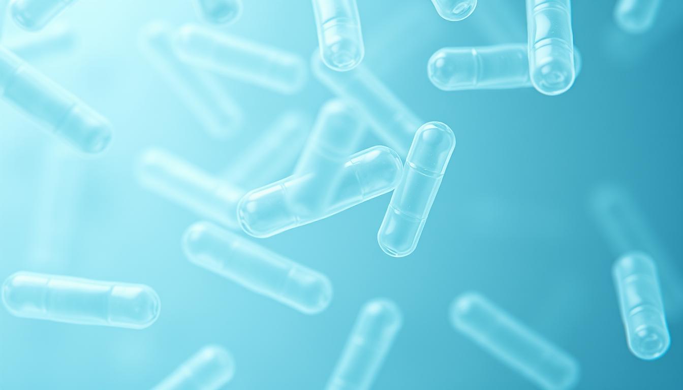
Gram-Stained E. coli: Characteristic Appearance
Looking at Gram-stained E. coli under a microscope shows clear features. It’s a Gram-negative, rod-shaped bacterium.
Typical Morphology Under Microscope
The shape of E. coli is key to identifying it. Under a microscope, it looks like rod-shaped cells.
Size and Shape Parameters
E. coli cells are 2.0-6.0 micrometers long and 1.1-1.5 micrometers wide. Their rod shape is a major identification feature.
Arrangement Patterns in Cultures
In cultures, E. coli cells can be alone or in pairs. This shows how they behave in different settings.
| Characteristic | Description |
|---|---|
| Shape | Rod-shaped |
| Size | 2.0-6.0 µm in length, 1.1-1.5 µm in width |
| Arrangement | Singles or pairs |
Interpreting the Pink/Red Coloration
The pink or red color in Gram-stained E. coli comes from safranin. This color is typical of Gram-negative bacteria.
Safranin Retention Mechanism
The safranin retention happens in the Gram staining process. Safranin stains Gram-negative cells pink or red.
Distinguishing True vs. False Gram-Negative Results
It’s important to tell true Gram-negative results from false negatives. False negatives can happen if the cells are too decolorized. Using the right technique is key for accurate results.
Cell Wall Structure of E. coli
Understanding E. coli’s cell wall is key to knowing it’s Gram-negative. The cell wall has several layers, each with its own role. These roles help the cell interact with its surroundings.
Outer Membrane Composition
The outer membrane of E. coli is complex and vital for its Gram-negative status. It’s made of a phospholipid bilayer and proteins. These proteins help with transport and signaling.
Phospholipid Bilayer Organization
The phospholipid bilayer gives the outer membrane its strength. The outer side has lipopolysaccharides, while the inner side has phospholipids.
Membrane Proteins and Their Functions
Membrane proteins in E. coli’s outer membrane do many things. They help move molecules across the membrane and act as receptors for signals.
Peptidoglycan Layer Characteristics
The peptidoglycan layer, or periplasmic peptidoglycan layer, adds strength to the cell wall. It’s thinner in E. coli than in Gram-positive bacteria.
Thickness and Structural Properties
The peptidoglycan layer in E. coli is about 2-3 nanometers thick. Its structure is important for keeping the cell’s shape and fighting off osmotic pressure.
Comparison with Gram-Positive Bacteria
E. coli’s peptidoglycan layer is much thinner than Gram-positive bacteria’s. This difference is why E. coli stains differently in the Gram staining technique.
Lipopolysaccharides and Their Function
Lipopolysaccharides (LPS) are key parts of E. coli’s outer membrane. They are important for how the bacterium interacts with its environment.
Endotoxin Properties
LPS are known for their ability to cause strong immune reactions. This is a big reason why some E. coli strains are harmful.
Role in Bacterial Survival
LPS help E. coli survive by protecting it from environmental stresses and the host’s immune system.
| Component | Function | Characteristics |
|---|---|---|
| Outer Membrane | Provides barrier function and contains lipopolysaccharides | Complex structure with phospholipid bilayer and proteins |
| Peptidoglycan Layer | Maintains cell shape and provides mechanical strength | Thin layer, approximately 2-3 nanometers thick |
| Lipopolysaccharides | Acts as endotoxin and contributes to bacterial survival | Complex molecules with lipid and polysaccharide components |
Step-by-Step E. coli Gram Staining Procedure
To identify E. coli, follow a detailed Gram staining procedure. This method is key in microbiology. It helps tell apart Gram-positive and Gram-negative bacteria.
Sample Preparation Techniques
Preparing the sample is the first and most important step. It includes choosing the right culture and preparing the smear.
Culture Selection and Handling
Picking the right E. coli culture is essential. It should be fresh and growing well. Handling must be done carefully to avoid contamination.
Smear Preparation Methods
Spread a small amount of culture on a clean slide to make a thin smear. Let it dry and then fix it with heat.
Primary Staining with Crystal Violet
The first step is applying crystal violet to the smear.
Application Technique
Spread the crystal violet stain evenly over the smear. Leave it for 1-2 minutes.
Timing Considerations
How long you leave the crystal violet on is important. Too long can cause overstaining.
Iodine Application as Mordant
After staining, apply iodine as a mordant. It helps fix the stain.
Function of Iodine in the Process
Iodine helps the crystal violet stain stick to the bacterial cell wall.
Proper Application Method
Apply iodine for about 1 minute. Make sure it covers the whole smear.
Decolorization Process
Decolorization is key to telling Gram-positive from Gram-negative bacteria.
Alcohol-Acetone Mixture Usage
Use an alcohol-acetone mix for decolorization. E. coli, being Gram-negative, will lose its stain.
Critical Timing Factors
How long you decolorize is very important. Too long can wash away the stain.
Counterstaining with Safranin
The last step is counterstaining with safranin.
Purpose of Counterstain
Safranin colors the Gram-negative bacteria pink or red. It acts as a counterstain.
Optimal Application Technique
Apply safranin for 1-2 minutes. Rinse gently with water and let dry.
As “The Gram stain is one of the most important staining techniques in microbiology”, following this guide ensures accurate E. coli identification.
Common Errors in E. coli Gram Staining
Getting E. coli Gram staining right is key in microbiology. Yet, many mistakes can mess up the results. The Gram stain technique for E. coli is a basic lab skill that needs careful attention.
Preparation Mistakes and Their Effects
Preparation errors are a big problem in E. coli Gram staining. Mistakes often happen in smear thickness and heat fixation.
Smear Thickness Issues
A too-thick smear can ruin the Gram staining. Thick smears pack bacteria too close, making it hard to see individual cells. On the other hand, a too-thin smear might not have enough bacteria for study.
Heat Fixation Problems
Heat fixation is key in getting E. coli samples ready for Gram staining. Not enough heat fixation can lose bacteria during staining. But too much heat can mess up the shape of the bacteria.
| Preparation Mistake | Effect on Gram Staining |
|---|---|
| Smear too thick | Inaccurate results due to over-concentration |
| Smear too thin | Insufficient bacteria for analysis |
| Insufficient heat fixation | Loss of bacteria during staining |
| Over-fixation | Distortion of bacterial morphology |
Staining Timing Issues
Timing is everything in the E. coli staining process. Mistakes in timing can give wrong results.
Overdecolorization Effects
Overdecolorization can make E. coli look Gram-positive instead of Gram-negative. This happens when the decolorizer stays on too long.
Inadequate Counterstaining
Not enough counterstaining with safranin can make bacteria hard to see. The counterstain is key for spotting Gram-negative bacteria like E. coli.
Knowing and avoiding these common mistakes helps lab techs get accurate E. coli Gram staining results.
Microscopy Techniques for Observing Stained E. coli
To study E. coli, scientists use advanced microscopy. This is key to understanding its shape and how it acts.
Light Microscopy Methods
Light microscopy is a basic way to see Gram-stained E. coli. It gives a clear look at the bacteria’s shape.
Brightfield Microscopy Settings
Brightfield microscopy is great for seeing stained bacteria. It’s important to set the condenser and light right for the best view.
Contrast Enhancement Techniques
Improving contrast is key for clear E. coli images. Adjusting the microscope and using special stains can make images better.
Oil Immersion Technique
The oil immersion technique is important for detailed E. coli images. It uses a special lens and oil to reduce light loss.
Proper Oil Application
Using the right amount of immersion oil is critical. Too little oil can cause poor contact, while too much can spread oil.
Magnification and Resolution Factors
The microscope’s magnification and resolution depend on several things. These include the lens’s numerical aperture and the light’s wavelength.
By using these microscopy methods, scientists can learn more about E. coli. This knowledge is vital for many microbiology uses.
Differentiating E. coli from Other Gram-Negative Bacteria
To tell E. coli apart from other Gram-negative bacteria, you need to know its unique traits. This is key in lab work, like in hospitals and when checking the environment.
Morphological Distinctions
E. coli stands out because of its shape. It’s a rod-shaped bacterium, about 2-3 μm long and 0.5-1 μm wide. When looking at E. coli and other Gram-negative bacteria, its shape is a big clue.
Comparison with Salmonella Species
Salmonella is also a Gram-negative rod, like E. coli. But Salmonella moves more, which you can see in wet mounts. Both are hard to break down lactose, but E. coli reacts more strongly in tests.
Distinguishing from Pseudomonas
Pseudomonas aeruginosa is known for its blue-green color on agar plates. It doesn’t break down lactose and is strictly air-breathing. Its colonies are bigger and stickier than E. coli‘s.
Additional Confirmatory Tests
Even with shape clues, more tests are needed for sure identification. These tests check how E. coli reacts biochemically and genetically.
Biochemical Test Panels
Tools like API or Enterotube systems check how bacteria react. E. coli breaks down lactose and makes indole, helping it stand out.
Molecular Identification Methods
Methods like PCR and 16S rRNA sequencing pinpoint E. coli accurately. They look for specific genes to tell E. coli from similar bacteria.
In summary, figuring out E. coli from other Gram-negative bacteria takes shape, biochemical, and genetic tests. Each method gives important info. Together, they ensure we identify E. coli correctly.
Clinical Significance of Identifying E. coli
Knowing how E. coli affects health is key to treating infections. E. coli is a type of bacteria found in our intestines. But, some strains can lead to serious infections.
Pathogenic vs. Non-Pathogenic Strains
E. coli can be divided into two main types: pathogenic and non-pathogenic. Pathogenic strains can cause disease. Non-pathogenic strains are safe and live in our gut.
EHEC, ETEC, and Other Pathotypes
Pathogenic E. coli includes types like EHEC and ETEC. EHEC can cause severe foodborne illness, leading to bloody diarrhea. ETEC is a common cause of traveler’s diarrhea.
Virulence Factors and Detection
Pathogenic E. coli has special features like adhesins and toxins. Finding these is important for spotting harmful strains.
| Characteristics | Pathogenic E. coli | Non-Pathogenic E. coli |
|---|---|---|
| Presence of Virulence Factors | Yes | No |
| Disease Causing Ability | Yes | No |
| Common Locations | Various, including intestine | Primarily intestine |
Role in Diagnostic Microbiology
E. coli is important in diagnosing infections. It helps in identifying urinary tract and gastrointestinal infections. Accurate identification is key for treatment.
Urinary Tract Infection Diagnosis
In UTIs, E. coli is a common culprit. Doctors isolate E. coli from urine and check its antibiotic resistance.
Gastrointestinal Infection Identification
E. coli also causes gastrointestinal infections. Knowing the type and its virulence is vital for treatment.
Environmental and Research Applications
E. coli’s impact goes beyond the lab. It’s key in environmental monitoring and molecular biology research. Its widespread use makes it perfect for many tasks.
E. coli as Environmental Indicator
E. coli in water means fecal contamination and health risks. This is vital for keeping us safe.
Water Quality Assessment
E. coli checks water quality well. It changes with the environment and shows pathogen presence. Monitoring E. coli levels ensures water is safe for us.
Fecal Contamination Monitoring
E. coli marks fecal contamination. It helps find sewage leaks in water. This stops waterborne disease outbreaks.
Research Uses in Molecular Biology
In molecular biology, E. coli is a key player. It’s used in genetic engineering and protein production.
Genetic Engineering Applications
E. coli is a top choice for genetic engineering. It’s easy to work with and has simple genetics. This makes it perfect for cloning and protein production.
Protein Expression Systems
E. coli is great for making proteins. It grows fast and produces lots of protein. This is very useful for research and medicine.
E. coli’s many uses show its value beyond the lab. It’s important in environmental monitoring and molecular biology. Its flexibility and importance are clear.
Advanced Staining Techniques for E. coli Visualization
Advanced staining techniques give us a deeper look at E. coli shape and how it acts. They show us more than just what Gram staining does.
Fluorescent Staining Methods
Fluorescent staining has changed how we see E. coli under the microscope. It lets us see special parts or traits of the cells.
DAPI and Nucleic Acid Stains
DAPI (4′,6-diamidino-2-phenylindole) is a special stain that sticks to DNA. It’s great for looking at the DNA in E. coli. Other stains like SYTO and SYBR dyes also help us understand if bacteria are alive and what their DNA is like.
Live/Dead Bacterial Viability Kits
Live/Dead kits use different colors to show if bacteria are alive or not. They’re really helpful in seeing if treatments work against E. coli.
Immunostaining Approaches
Immunostaining is a precise way to spot E. coli in complicated samples.
Antibody-Based Detection Systems
Antibody-based systems use special antibodies made for E. coli. These antibodies can glow, helping us find E. coli cells.
Fluorescent Antibody Techniques
Fluorescent antibody methods use glowing antibodies to find certain E. coli parts. This is great for telling E. coli apart from other bacteria in mixtures.
| Staining Technique | Application | Key Features |
|---|---|---|
| DAPI Staining | Nucleic acid visualization | Binds to DNA, fluorescent |
| Live/Dead Viability Kits | Viability assessment | Differentiates viable and non-viable cells |
| Fluorescent Antibody Techniques | Specific antigen detection | High specificity, fluorescent labeling |
These advanced staining methods have greatly improved our study of E. coli and other bacteria. They give us important details about their shape, behavior, and how they interact.
Digital Imaging and Analysis of Gram-Stained E. coli
The way we look at E. coli has changed a lot with new digital imaging. This makes it easier and more accurate to study Gram-stained E. coli.
Modern Imaging Technologies
New imaging tech has changed microbiology a lot, mainly in studying Gram-stained E. coli. Digital microscopy systems are now key in research and testing.
Digital Microscopy Systems
These systems give us clear, detailed images. They let us capture, save, and study images digitally.
Image Capture Optimization
Getting the best images of Gram-stained E. coli is key. Adjusting light and focus are important steps.
Quantitative Analysis Methods
There are many ways to analyze Gram-stained E. coli. These methods help us get useful data from images.
Automated Cell Counting Software
Software for counting cells automatically makes research faster and more precise. It cuts down on manual work.
Morphometric Analysis Tools
Tools for morphometric analysis let us closely look at E. coli’s shape and size. This is very useful.
| Analysis Method | Description | Application |
|---|---|---|
| Automated Cell Counting | Software that counts E. coli cells automatically | Quantifying bacterial load |
| Morphometric Analysis | Tools that measure E. coli cell dimensions | Studying morphological changes |
| Digital Microscopy | Systems that capture high-resolution images | Enhancing image quality |
In conclusion, digital imaging and analysis have greatly improved our study of Gram-stained E. coli. They make E. coli morphology analysis more accurate and efficient.
Conclusion
Understanding Gram-Stained E. coli is key in microbiology. It’s a model organism used widely. The E. coli staining procedure uses Gram staining. This method sorts bacteria into Gram-positive and Gram-negative groups based on their cell walls.
Gram-Stained E. coli looks pink or red and rod-shaped under a microscope. This is because of its Gram-negative cell wall. The staining process includes primary staining with crystal violet, decolorization, and counterstaining with safranin.
Identifying Gram-negative bacteria like E. coli is important in many areas. This includes clinical diagnostics, environmental monitoring, and research. Accurate identification depends on Gram staining and other tests.
In summary, studying Gram-Stained E. coli is essential. It helps us understand its role in different fields. By learning the E. coli staining procedure and Gram staining, researchers and clinicians can better identify and analyze E. coli. This contributes to progress in microbiology and related fields.