The Neurenteric Canal is key in embryonic development. It forms early in an embryo’s growth.
Problems with this canal can affect an embryo’s growth. Knowing about the Neurenteric Canal helps us understand embryonic development. It also shows how issues with it can lead to birth defects.
The growth of an embryo is complex. The formation of the Neurenteric Canal is a vital part of it. Issues with its formation can have big effects.
The Neurenteric Canal: Definition and Overview
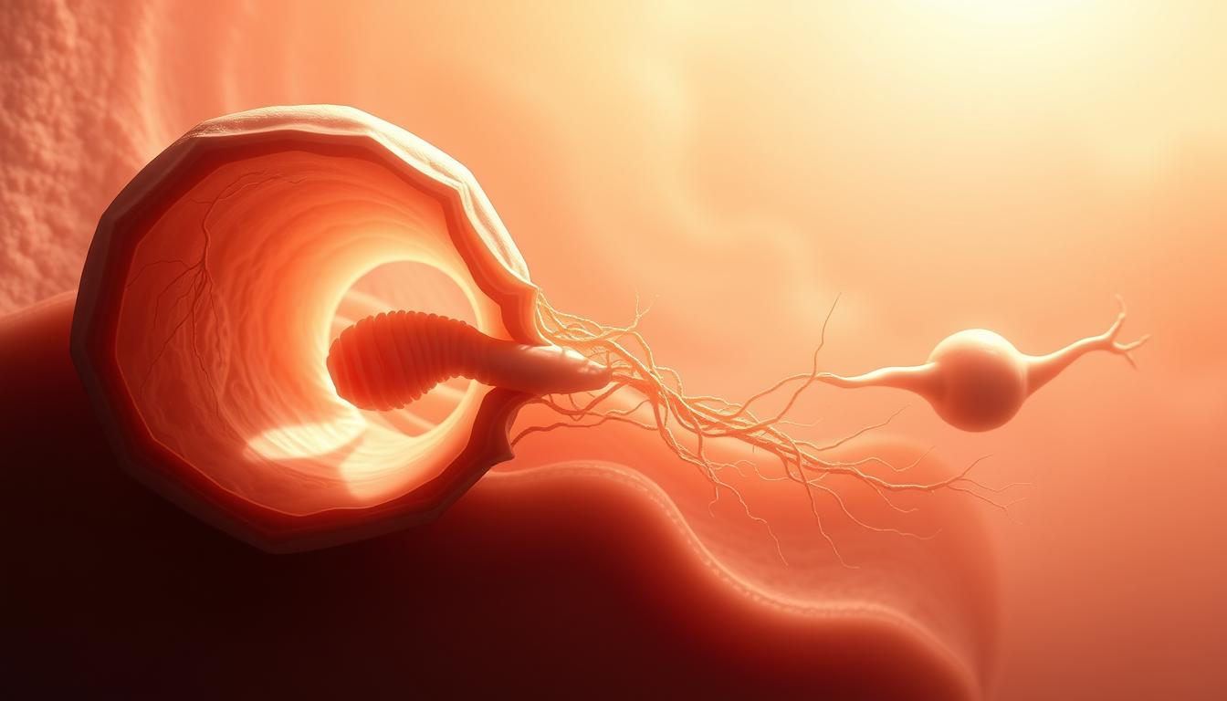
The Neurenteric Canal is a key link between the neural tube and the early gut during development.
This canal is a temporary structure that is vital in the early stages of growth. It connects the neural tube, which forms the brain and spinal cord, to the primitive gut. This gut will eventually become the digestive system.
Historical Context and Discovery
The Neurenteric Canal was first found in early studies of embryos. It was seen as a temporary link between the yolk sac or early gut and the amniotic cavity. Early embryologists found it in many species, showing its importance in developmental biology.
| Year | Researcher | Contribution |
|---|---|---|
| 1850 | Johann Friedrich Meckel | Initial descriptions of the Neurenteric Canal |
| 1920 | Experimental embryologists | Detailed its role in embryonic development |
Anatomical Positioning in the Embryo
The Neurenteric Canal is found between the neural tube and the early gut. It is a transient structure that helps in the exchange of cells and signals. Its location is close to the notochord, which is important for the development of the spinal column.
Embryological Origins of the Neurenteric Canal
Gastrulation is a key phase in early development. It sets the stage for the Neurenteric Canal’s creation. This stage involves cells moving together to form the ectoderm, endoderm, and mesoderm layers.
Gastrulation and Early Embryonic Development
During gastrulation, the blastula transforms through detailed cell movements. This change is vital for the embryo’s growth and the creation of the Neurenteric Canal. Cells from the primitive node move forward to form the notochordal process.
The notochordal process plays a big role in the Neurenteric Canal’s development. It helps create the neural plate and shapes the surrounding tissues.
Timeline of Neurenteric Canal Formation
The Neurenteric Canal starts to form in the third week of development. This is a time of fast growth and cell specialization. Its formation is closely linked to gastrulation and the notochord’s development.
| Developmental Stage | Timeline | Description |
|---|---|---|
| Gastrulation | Week 3 | Formation of the three primary germ layers |
| Notochordal Process Formation | Week 3 | Development of the notochordal process |
| Neurenteric Canal Formation | Week 3 | Emergence of the Neurenteric Canal |
The exact timing of the Neurenteric Canal’s formation highlights its importance in early development.
The Process of Neurenteric Canal Formation
The Neurenteric Canal forms through complex molecular steps. This is key for the embryo’s growth, linking the neural and enteric systems.
This canal starts to form early in development, during gastrulation. This stage sees big changes in cells and their roles. It sets the stage for organs to develop later.
Molecular Mechanisms
Many molecules work together to create the Neurenteric Canal. Key players include signaling molecules and transcription factors. They guide the canal’s formation.
- Sonic Hedgehog (SHH) and Wnt/β-catenin help shape the canal.
- Brachyury and Sox2 are vital for its development.
A study found that these molecules work together. They ensure the Neurenteric Canal forms correctly. This is essential for the embryo’s growth.
“The Neurenteric Canal is a transient structure that plays a critical role in the embryogenesis of the nervous system and the gastrointestinal tract.”
Key Developmental Signaling Pathways
Several pathways are key to the Neurenteric Canal’s formation. These include:
| Signaling Pathway | Role in Neurenteric Canal Formation |
|---|---|
| SHH Signaling | Regulates patterning and development of the canal |
| Wnt/β-catenin Signaling | Involved in the regulation of cellular differentiation and proliferation |
| Notch Signaling | Plays a role in cell fate decisions and boundary formation |
The complex interaction of these pathways is vital. It ensures the Neurenteric Canal forms and then disappears properly. This is essential for the embryo’s development.
Structural Characteristics of the Neurenteric Canal
The Neurenteric Canal is a key structure in early development. It has unique features that help us understand its importance. Knowing about its cells and how it changes helps us see its role in growth.
Cellular Composition
The Neurenteric Canal is made up of different cell types. Epithelial cells are at the center, playing a big role in its creation and upkeep. These cells have distinct morphology and molecular markers that set them apart from other cells in the embryo.
As the embryo grows, the Neurenteric Canal’s cells change a lot. These cells work with the tissues around them, helping other parts of the embryo develop.
| Cell Type | Characteristics | Role in Neurenteric Canal |
|---|---|---|
| Epithelial Cells | Distinct morphology, molecular markers | Formation and maintenance |
| Mesenchymal Cells | Supportive, migratory capabilities | Structural support, interaction with surrounding tissues |
Dimensional Changes During Development
The Neurenteric Canal changes a lot as the embryo grows. At first, it’s big, helping the neural and enteric systems talk to each other. But as growth goes on, it gets smaller and eventually disappears.
The regression of the Neurenteric Canal is a complex process, influenced by a multitude of genetic and environmental factors.
It’s important to understand these changes to see how the canal affects development. Knowing this helps us understand what happens if it doesn’t close properly.
The changes in the Neurenteric Canal are tied to the embryo’s overall growth. If these changes don’t happen right, it can cause big problems in development.
Functional Significance in Embryonic Development
The Neurenteric Canal is key in early development, helping different parts of the embryo talk to each other. It’s only there briefly but has a big impact. It helps the neural and enteric systems work together and aids in the growth of the notochord.
Communication Between Neural and Enteric Systems
The Neurenteric Canal connects the neural tube and the early gut. This connection is vital for their proper growth. Without it, these systems might not develop right.
It’s all about the molecular signals. These signals help cells in both systems grow and organize correctly. The Neurenteric Canal’s role shows its importance in early development.
Role in Notochord Development
The Neurenteric Canal also plays a part in the notochord’s growth. The notochord is a key structure that helps form the spine and the brain. Any issues here can cause problems with the notochord.
The connection between the Neurenteric Canal and the notochord shows how complex early development is. Understanding this relationship helps us learn more about how embryos grow.
| Aspect | Description | Implications |
|---|---|---|
| Communication Between Neural and Enteric Systems | Facilitates signal exchange between neural tube and primitive gut | Crucial for coordinated development of nervous system and gastrointestinal tract |
| Role in Notochord Development | Involved in the development of the notochord | Impacts vertebral column formation and central nervous system development |
Normal Regression of the Neurenteric Canal
The Neurenteric Canal’s regression is key in early development. It’s vital for the right growth of the embryo’s neural and enteric systems.
Timing of Regression
The Neurenteric Canal’s regression happens at a set time in development. Studies have shown it usually occurs between the third and fourth week of pregnancy in humans. This is when the embryo goes through major changes, and the canal’s regression is a critical event.
The exact timing can vary a bit, but it’s mostly the same for everyone. Knowing this timeline helps spot any issues in development.
Cellular Mechanisms of Canal Closure
The way the Neurenteric Canal closes is complex. Cellular proliferation, differentiation, and apoptosis are all important. As the canal closes, the cells around it change in specific ways to help it shut down.
- Cellular proliferation helps fill the canal.
- Apoptosis, or programmed cell death, helps it close.
- Differential growth and morphogenetic movements also play a role.
These steps work together to close the Neurenteric Canal properly. This is essential for normal development. Any problems can cause developmental issues.
Relationship to Notochord Development
Understanding the link between the Neurenteric Canal and notochord development is key. The Neurenteric Canal connects the yolk sac to the amniotic cavity early in development. It plays a big role in how embryos grow.
Notochord Formation and the Neurenteric Canal
The notochord is a key part of the spinal column and helps form the central nervous system. The Neurenteric Canal is important for notochord development. Abnormalities in the Neurenteric Canal can cause notochord problems, leading to spinal issues.
Creating the notochord is a complex process. It involves cells moving and signaling pathways. The Neurenteric Canal’s role shows its importance in early development.
Implications for Spinal Development
The connection between the Neurenteric Canal and the notochord is very important for the spine. Notochord abnormalities can cause spinal defects like spina bifida. Knowing about this connection helps doctors diagnose and treat spinal problems.
Also, the Neurenteric Canal’s disappearance is vital for the spine to develop right. If it doesn’t close, it can cause problems. This can lead to spinal and brain issues.
Neurenteric Canal Abnormalities and Clinical Presentations
The neurenteric canal is a temporary part of early development. It can have problems that affect growth and health later on.
Persistent Neurenteric Canal
A persistent neurenteric canal is a rare birth defect. It happens when the canal doesn’t close up as it should. This can cause problems like digestive and brain issues.
Doctors use MRI or CT scans to find this problem. Surgery might be needed to fix it and make symptoms better.
Neurenteric Cysts
Neurenteric cysts are benign growths from the neurenteric canal. They are filled with fluid and can appear in different parts of the body, like the chest or spine.
These cysts can press on nearby tissues, causing symptoms. Removing the cyst through surgery is usually the treatment to stop further problems.
Gastrointestinal Abnormalities Related to Neurenteric Canal Defects
The Neurenteric Canal is key in early development. Its defects can cause big problems with the gut. If it doesn’t close up right, it can lead to many gut issues.
Duodenal Stenosis and Intestinal Malformations
Duodenal stenosis is a big problem. It makes the duodenum narrow, causing blockages. Other issues like malrotation and atresia can also happen because of the gut’s wrong development.
The reason for these problems is tied to how the Neurenteric Canal forms. If it doesn’t close up right, it messes with the gut’s growth. This can cause these issues.
- Duodenal stenosis
- Intestinal malrotation
- Intestinal atresia
Diagnostic Approaches and Management
Figuring out gut problems linked to Neurenteric Canal defects needs special tests. Prenatal ultrasound can spot some issues. But MRI and CT scans give a clearer picture after birth.
Handling these problems needs a team effort. Doctors, surgeons, and radiologists all play a part. Surgery is often needed to fix things, like opening up the duodenum.
Neurological Disorders Associated with Neurenteric Canal Anomalies
Abnormalities in the Neurenteric Canal are linked to many neurological disorders. This shows how important it is for the canal to form correctly. The Neurenteric Canal is key in early development, and problems with it can cause serious neurological issues.
Neuraxis Development Disorders
Neuraxis development disorders are a big worry with Neurenteric Canal issues. The neuraxis, or central nervous system, can be harmed by problems with the Neurenteric Canal. Disorders of neuraxis development can cause problems with movement and thinking.
The neuraxis develops through a complex mix of genetics and environment. Disruptions in this process can cause problems with the central nervous system’s structure and function.
Hindbrain Deformities and Clinical Implications
Hindbrain deformities are another issue linked to Neurenteric Canal problems. The hindbrain, which includes the cerebellum and brainstem, is vital for controlling important functions and movement. Deformities in this area can cause big problems, like balance and coordination issues.
The effects of hindbrain deformities can vary a lot. Early diagnosis and management are key to reducing these problems and helping patients.
In summary, Neurenteric Canal issues can cause many neurological problems, like neuraxis and hindbrain issues. Knowing about these connections is vital for diagnosing and treating related conditions effectively.
Diagnostic Imaging of Neurenteric Canal Abnormalities
Diagnostic imaging is key in spotting Neurenteric Canal problems. It uses advanced methods for precise diagnosis and treatment plans.
Prenatal Ultrasound Findings
Prenatal ultrasound is vital for catching Neurenteric Canal issues early. It checks for possible problems and helps decide on pregnancy care.
Ultrasound might show cysts or oddities in the gut. These signs could point to Neurenteric Canal defects.
MRI and CT Evaluation
MRI and CT scans give detailed pictures for checking Neurenteric Canal issues. MRI is great for soft tissues, while CT is better for bones.
These scans help figure out how serious the problem is. They also help plan surgery if needed.
Differential Diagnosis Considerations
When looking at Neurenteric Canal issues, other possible problems must be considered. Things like enteric duplication cysts or other gut malformations need to be ruled out.
A detailed diagnostic process is needed. This includes imaging and clinical checks to get a correct diagnosis.
The right imaging choice depends on several things. These include the fetus’s age, the suspected issue, and the symptoms.
Surgical Management of Neurenteric Canal Anomalies
Fixing Neurenteric Canal anomalies needs a team effort from different doctors. These problems are complex and need a treatment plan made just for each patient.
Pediatric Surgical Approaches
Pediatric surgeons use careful methods to fix these anomalies. They aim to avoid harming nearby tissues. These patients will need ongoing care after surgery.
The right surgery depends on the anomaly’s details. Doctors use advanced imaging to plan the surgery well.
Multidisciplinary Team Involvement
A team of doctors is key to treating Neurenteric Canal anomalies. This team includes pediatric surgeons, neurosurgeons, and radiologists. They work together to create a detailed treatment plan.
Postoperative Care and Long-term Outcomes
After surgery, patients are closely watched for any problems. Their long-term health depends on the surgery’s success and the anomaly’s severity.
| Surgical Approach | Advantages | Disadvantages |
|---|---|---|
| Minimally Invasive Surgery | Less tissue damage, quicker recovery | Limited visibility, technical challenges |
| Open Surgery | Better visualization, more control | More tissue damage, longer recovery |
The table shows the good and bad sides of different surgeries for Neurenteric Canal anomalies. The choice of surgery depends on the patient’s health and the anomaly’s details.
Recent Advances in Understanding Neurenteric Canal Development
New discoveries in embryology have greatly improved our understanding of the Neurenteric Canal. These findings have shown how this canal forms and then disappears. This knowledge is key to understanding its role in development.
Genetic Studies and Molecular Insights
Recent studies have found important genes and pathways for the Neurenteric Canal. For example, certain transcription factors control cell actions during growth. The Wnt/β-catenin pathway is also vital for cell development.
- Research has shown how genetic mutations can affect the canal’s development.
- There’s interest in molecular diagnostics for early detection of issues.
Animal Models and Translational Research Findings
Mouse and chick embryos have been key in studying the Neurenteric Canal. These models let researchers change genetic and environmental factors to see their effects. The findings from these studies help us understand human issues and find new treatments.
- CRISPR-Cas9 gene editing in animals has made it easier to study specific genes.
- Preclinical studies in animals are essential for applying research to humans.
These recent breakthroughs show how complex the Neurenteric Canal’s development is. They highlight the need for ongoing research into its genetic and molecular aspects.
Conclusion
The Neurenteric Canal is key in early development, linking the brain and gut systems. It’s important for the embryo’s growth. Its creation and disappearance are vital steps.
Problems with the Neurenteric Canal can cause serious health issues. This includes problems with the gut and brain. Knowing how it forms helps doctors diagnose and treat related birth defects.
Understanding the Neurenteric Canal’s role in development is critical. More research is needed to learn about its impact on health. This will help us better understand its role in human health and disease.