The Kennedy Terminal Ulcer is a specific skin condition often seen in end-of-life care. It’s important for healthcare professionals to understand this condition well.
This condition has a unique presentation. It needs to be identified and managed correctly to ensure patient comfort. The importance of recognizing the Kennedy Terminal Ulcer is huge for patient care.
By knowing the characteristics of this condition, healthcare providers can manage it better. This helps improve patient outcomes. It’s key to have the right care and management strategies for patients with this condition.
The Clinical Significance of Kennedy Terminal Ulcers
Kennedy Terminal Ulcers are special because of how they look and the challenges they bring. They are a type of skin problem that happens to people who are very sick.
Definition and Distinctive Features
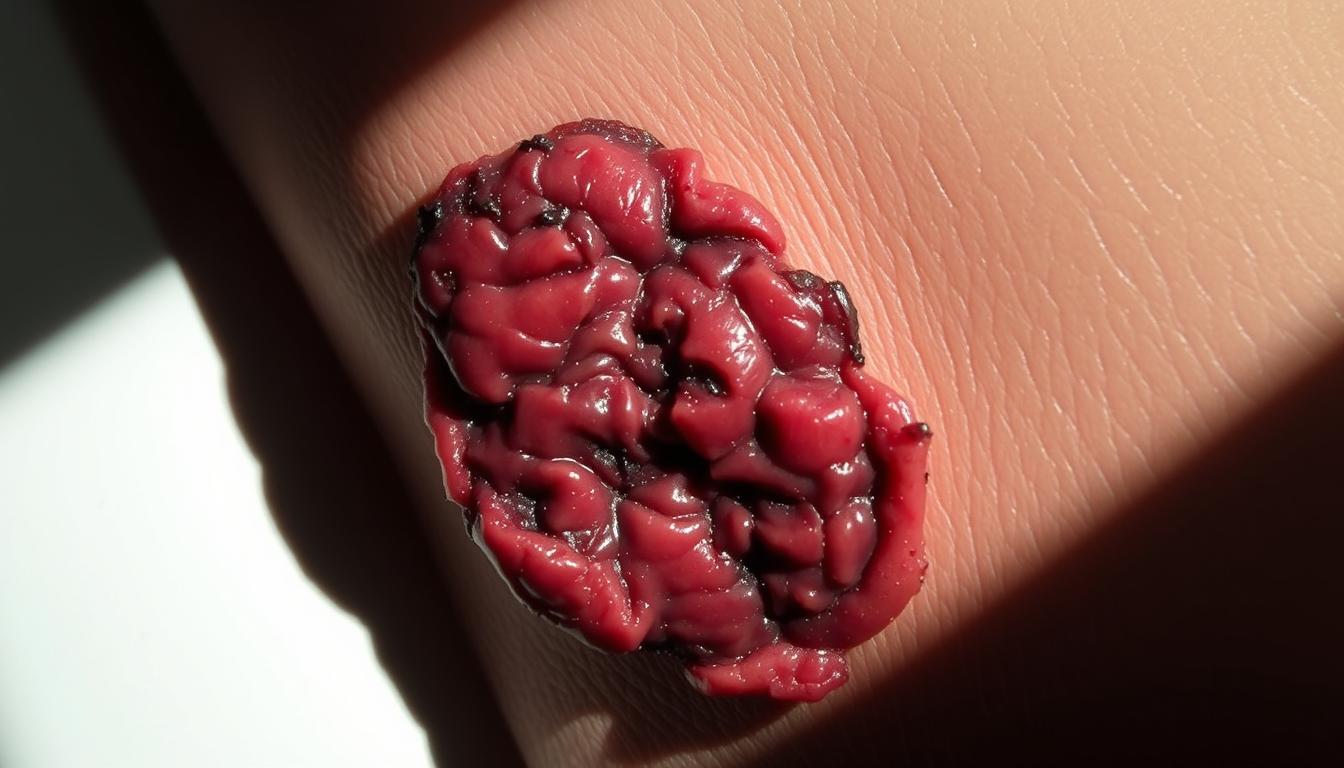
Kennedy Terminal Ulcers start quickly and have a unique shape, often looking like a “pear” or “butterfly.” They usually appear on the sacrum or coccyx. Their growth is tied to the patient’s health and how much they can move.
Karen Lou Kennedy says these ulcers show the body’s shutdown. Experts in wound care agree, saying they show the body’s failure to keep things stable.
Prevalence and Impact on Patient Care
Kennedy Terminal Ulcers are common in end-of-life care, affecting many patients in hospice. They make patient care harder, needing changes in how wounds are managed and care for comfort. Doctors and nurses must understand how these ulcers affect patients’ comfort and life quality.
Knowing about Kennedy Terminal Ulcers is key to giving good care to those with these tough wounds. By understanding their unique signs and what they mean, healthcare workers can improve care and results.
Historical Background of the Kennedy Ulcer
The Kennedy Ulcer has a rich history tied to end-of-life care. It’s a condition closely linked to decubitus ulcer. It has drawn a lot of attention in medical settings because of its unique traits and the care it requires.
Karen Lou Kennedy’s Original Observations
Karen Lou Kennedy’s work was key in spotting the Kennedy Ulcer as a unique condition. Her findings helped us understand this tissue injury better. Kennedy’s work showed how it differs from other skin problems.
Her research shed light on the special needs of patients with this condition, mainly in end-of-life care. Recognizing the Kennedy Ulcer as its own entity has changed how we treat and care for these patients.
Evolution of Clinical Understanding
After Karen Lou Kennedy’s early findings, our understanding of the Kennedy Ulcer has grown a lot. More studies have helped us learn about its causes, risk factors, and how to manage it. Now, the medical field sees the Kennedy Ulcer as a complex issue, often linked to the dying process.
This deeper understanding has led to better ways to diagnose and treat Kennedy Ulcers. It has improved the care for those with this condition. Ongoing research keeps helping us provide better, more compassionate care to these patients.
Decoding the “3:30 Syndrome” Phenomenon
Exploring the “3:30 Syndrome” linked to Kennedy Terminal Ulcers offers valuable insights. This phenomenon is a key feature of Kennedy Terminal Ulcers. It has caught the attention of the wound care field because of its unique characteristics.
Origin and Meaning of the Term
The term “3:30 Syndrome” comes from the typical look of Kennedy Terminal Ulcers. They often look like the time 3:30 on a clock. This shape is not just a coincidence but is tied to the ulcers’ underlying cause.
The “3:30 Syndrome” is more than just a name; it points to a specific way ulcers grow. This pattern is connected to the body’s changes in patients with Kennedy Terminal Ulcers.
Characteristic Butterfly or Pear-Shaped Appearance
Kennedy Terminal Ulcers often look like a butterfly or a pear. This unique shape is a key sign that helps doctors tell them apart from other skin ulcers. The shape comes from how pressure and lack of oxygen affect the skin.
Rapid Onset and Development Pattern
The “3:30 Syndrome” is known for its quick start and growth. Kennedy Terminal Ulcers can grow fast, sometimes in just hours or days. Quick action is key to handle these ulcers well and help patients.
The fast growth of Kennedy Terminal Ulcers highlights the importance of watching patients closely. This is true for those at high risk.
Unilateral Presentation: A Hallmark Characteristic
Kennedy Terminal Ulcers often appear on one side of the body. This makes them tricky to diagnose and treat. Knowing about this unique presentation is key to spotting Kennedy Terminal Ulcers.
Anatomical Distribution Patterns
Kennedy Terminal Ulcers follow certain patterns in where they appear. They usually show up in areas that get a lot of pressure, like the sacrum, heels, or buttocks.
- Sacral area: A common site due to prolonged supine positioning.
- Heels: Frequently affected in patients with limited mobility.
- Buttocks: Areas over the ischial tuberosities are also prone to ulcer development.
Common Sites of Unilateral Development
Kennedy Terminal Ulcers often appear on one side of the body. This side is usually where the patient lies or sits. The unilateral development depends on how the patient moves, their health, and where they sit or lie.
Bilateral vs. Unilateral Manifestations
Even though Kennedy Terminal Ulcers usually show up on one side, they can also appear on both sides. It’s important to know the difference between these two types. Bilateral ulcers might mean there’s a bigger problem with how pressure is distributed and skin health.
In summary, the one-sided appearance of Kennedy Terminal Ulcers is a big clue for doctors. By understanding where these ulcers tend to appear, doctors can catch them early and treat them better.
Pathophysiology of Kennedy Terminal Ulcers
To understand Kennedy Terminal Ulcers, we must look at how tissue injury and skin breakdown are linked. These ulcers aren’t just caused by pressure. They come from a mix of factors that lead to their formation.
Skin Perfusion and Tissue Hypoxia
Skin perfusion is key to keeping tissues healthy. In Kennedy Terminal Ulcer patients, poor skin perfusion causes tissue hypoxia. This means tissues don’t get enough oxygen.
Tissue hypoxia is a major reason for these ulcers. Without enough oxygen, cells die, and ulcers form. Knowing this helps us find better ways to manage them.
Vascular Compromise Mechanisms
Vascular compromise is also important in Kennedy Terminal Ulcers. Things like heart problems, artery disease, and blockages can reduce blood flow. This makes tissue hypoxia worse.
The vascular compromise can come from many sources. It could be heart issues or pressure on blood vessels. Spotting these can help us prevent ulcers in some patients.
Cellular and Molecular Changes
At a cellular and molecular level, Kennedy Terminal Ulcers show inflammation, oxidative stress, and poor wound healing. The body’s response to lack of oxygen and blood flow involves many chemicals. These can either help or hinder healing.
Grasping these cellular and molecular changes is vital. It helps us create treatments that can change the disease’s course and improve patient results.
Differentiating Kennedy Ulcers from Other Pressure Injuries
It’s key to tell Kennedy Ulcers apart from other pressure injuries for better patient care. Kennedy Ulcers have unique traits that make them different from usual pressure ulcers and skin damage from moisture.
Comparison with Traditional Pressure Ulcers
Kennedy Ulcers look and grow differently than traditional pressure ulcers. They often show up as a one-sided, pear-shaped or butterfly-shaped sore.
Key differences include:
- Rapid onset and development
- Unilateral presentation
- Distinctive shape and appearance
Distinction from Moisture-Associated Skin Damage
MASD can look like Kennedy Ulcers, but it’s caused by long-term moisture exposure. This leads to skin irritation and damage.
| Characteristics | Kennedy Ulcers | MASD |
|---|---|---|
| Cause | Not directly related to moisture | Prolonged moisture exposure |
| Appearance | Pear-shaped or butterfly-shaped | Irregular, diffuse damage |
| Location | Typically unilateral, often on sacrum or heels | Variable, often in skin folds |
Diagnostic Challenges in Clinical Practice
It’s hard to diagnose Kennedy Ulcers because they look like other skin problems. Doctors need to know the special signs of Kennedy Ulcers to make the right diagnosis.
Risk Factors and Vulnerable Populations
It’s important to know the risk factors and who is most at risk for Kennedy Terminal Ulcers. Patients nearing the end of life are very vulnerable. This is because of changes in their body, other health issues, and how their body processes things.
End-of-Life Physiological Changes
As life comes to an end, patients face many changes that make them more likely to get skin ulcers. These changes include moving less, having poor blood flow, and skin that’s not as strong. Poor skin perfusion can cause tissues to not get enough oxygen, making the skin more prone to damage.
A study showed that patients who can’t move much are more likely to get skin ulcers, including Kennedy Terminal Ulcers. This highlights the need for moving them regularly and using strategies to avoid pressure on their skin.
Comorbidities and Predisposing Conditions
Some health conditions can make it more likely for someone to get Kennedy Terminal Ulcers. These include heart disease, diabetes, and neurological disorders. Diabetes, for example, can slow down wound healing because it can damage nerves and blood vessels.
| Comorbidity | Risk Factor | Impact on Skin |
|---|---|---|
| Cardiovascular Disease | Poor perfusion | Increased risk of skin ulcers |
| Diabetes | Neuropathy and vascular issues | Impaired wound healing |
| Neurological Disorders | Immobility and sensory loss | Increased pressure on skin |
Nutritional and Metabolic Factors
Nutritional deficiencies and changes in how the body uses nutrients can also affect the risk of getting Kennedy Terminal Ulcers. Eating well is key to keeping the skin healthy and helping wounds heal. Malnutrition can make it harder for the body to make collagen and repair tissues.
A study on wound care said something important about nutrition:
“Optimal nutrition is key for wound healing, as it gives the body what it needs to fix and grow new tissue.”
In summary, knowing the risk factors and who is most at risk is vital for preventing and managing Kennedy Terminal Ulcers. Healthcare workers need to watch for these signs to take the right steps to prevent them.
Assessment and Staging of Kennedy Ulcers
Understanding Kennedy Ulcers starts with a detailed assessment. This is key for the right care and management. The process includes several important steps to diagnose and stage these ulcers accurately.
Clinical Evaluation Techniques
Assessing Kennedy Ulcers involves a close look and checking the ulcer’s size, depth, and look. It’s also important to consider the patient’s overall health. This includes their mobility and nutrition, which can affect the ulcer’s growth.
Visual inspection is very important. It helps doctors spot the typical “pear-shaped” or “butterfly” look of Kennedy Ulcers. Using standard tools can make the evaluation more precise.
Documentation and Monitoring Protocols
Keeping accurate documentation is vital for tracking Kennedy Ulcer progress and checking treatment success. Doctors should write down all the details about the ulcer, like its size and look, and any changes over time.
Regular checks are key to catching any changes in the ulcer early. This might mean doctors seeing patients often and using photos or other ways to track changes.
Imaging and Advanced Assessment Methods
In some cases, imaging techniques like ultrasound or thermography are used. They help see how much tissue is damaged or how well it’s healing. These methods give extra info that helps doctors plan better.
Using advanced methods helps doctors understand the ulcer better. By combining these with clinical checks, they can make treatment plans that work better. This leads to better results for patients.
Comprehensive Management Approaches
Managing Kennedy Terminal Ulcers well needs a mix of wound care, pain management, and teamwork. This approach is key to better patient results and care quality.
Wound Care Principles and Dressing Selection
Starting with the right wound care is essential. Choosing the best dressing is vital for healing and comfort. Dressings that keep the wound moist and manage fluid are best. Advanced wound dressings like foam or hydrofiber work well for these ulcers.
Pain Management Strategies
Pain control is a big part of caring for Kennedy Terminal Ulcer patients. Assessing pain regularly is important because the pain can be severe. Treatment options include medicines and non-medical methods like changing the dressing to reduce pain.
Interdisciplinary Team Involvement
Having a team of experts helps a lot in treating Kennedy Terminal Ulcers. This team might include wound specialists, nurses, doctors, and others. They work together to create a detailed care plan. This ensures all aspects of care are covered, from wound treatment to pain relief.
Using a complete management plan, healthcare teams can improve care for Kennedy Terminal Ulcer patients. This leads to better comfort and results for them.
Preventive Measures in High-Risk Patients
To stop Kennedy Terminal Ulcers in people at risk, we need to act early. This means spotting risk factors, using special mattresses, and taking good care of the skin.
Risk Assessment Tools and Protocols
It’s important to use special tools to find out who might get Kennedy Terminal Ulcers. These tools help us catch problems early and act fast.
Common risk assessment tools include:
- The Braden Scale
- The Norton Scale
Positioning and Pressure Redistribution
Changing a person’s position often and using special mattresses are key. They help avoid too much pressure on sensitive spots.
Effective repositioning techniques involve:
- Turning patients regularly
- Using pillows or wedges to support positioning
| Preventive Measure | Description | Benefits |
|---|---|---|
| Risk Assessment Tools | Standardized scales to assess patient risk | Early identification, targeted intervention |
| Pressure Redistribution | Frequent turning, support surfaces | Reduced pressure on vulnerable areas |
| Skin Care | Gentle cleansing, moisturizing | Maintained skin integrity |
Skin Care and Integrity Maintenance
Keeping the skin clean and moisturized is key. Stay away from harsh soaps and use barrier creams to protect the skin.
Ethical Considerations in End-of-Life Skin Care
Managing skin integrity at the end of life raises big ethical questions. Healthcare workers must find a balance. They need to prevent or treat skin problems without hurting the patient’s comfort or dignity.
Balancing Interventions with Comfort Care
When life is ending, care shifts to comfort and relief. This means looking closely at the good and bad of skin care actions. For example, turning a patient to avoid decubitus ulcers might be painful, raising questions about comfort versus preventing harm.
Healthcare providers must think hard about the benefits and risks of actions. They need to understand the patient’s situation, values, and wishes for end-of-life care.
Family Education and Decision-Making Support
Family members are key in making decisions for patients nearing the end of life. It’s important to teach them about the ethics of skin care. This includes the good and bad of different treatments.
Supporting families is also vital. They might feel sad or unsure about their choices. Healthcare workers can offer help and guidance. This ensures care is in the patient’s best interest.
Recent Advances in Kennedy Ulcer Research and Treatment
The field of wound care has seen big steps forward in understanding Kennedy Ulcers. New studies aim to better treat and predict this condition.
Emerging Treatment Modalities
New ways to treat Kennedy Ulcers are being looked into. These include advanced dressings and new therapies. For example, negative pressure wound therapy is showing great promise in healing wounds.
| Treatment Modality | Description | Benefits |
|---|---|---|
| Negative Pressure Wound Therapy | Application of negative pressure to enhance wound healing | Promotes granulation tissue formation, reduces bacterial load |
| Advanced Wound Dressings | Use of dressings with antimicrobial properties | Reduces infection risk, maintains moist wound environment |
Biomarkers and Predictive Indicators
Scientists are working hard to find biomarkers for Kennedy Ulcer prediction. They’re looking at inflammatory markers and tissue perfusion. Finding these biomarkers could lead to better treatment and outcomes.
Ongoing Clinical Studies and Future Directions
Many clinical studies are exploring new treatments for Kennedy Ulcers. They aim to give doctors clear guidelines and improve care. Future research might focus on creating treatment plans tailored to each patient.
Conclusion
The Kennedy Terminal Ulcer, also known as the “3:30 syndrome,” is a special pressure sore. It shows up in patients close to the end of their life. It’s key for healthcare workers to know about this to give the best care.
Kennedy Ulcers look different from regular pressure ulcers. They have a butterfly or pear shape. Spotting these ulcers is important for correct diagnosis and treatment.
Managing Kennedy Ulcers well means using a full approach. This includes caring for the wound, managing pain, and working together as a team. It’s also important to prevent these ulcers in high-risk patients.
New research is helping us find better treatments and markers for Kennedy Ulcers. More study on this topic will help us understand and treat it better.
By understanding and treating Kennedy Terminal Ulcers well, healthcare teams can improve patient care. This leads to a better quality of life for those with this condition.