Knowing the head’s complex anatomy is key for healthcare workers. Using precise terms from ancient Greek and Latin makes communication clearer.
In the normal anatomical position, we use terms like “occipital” to identify body parts. This helps us accurately describe body structures. Understanding Head Zones is essential for mapping the body and pinpointing specific areas.
By diving into the different Head Zones and their importance, healthcare pros can boost their knowledge. This helps them give better care to their patients.
The Fundamental Concept of Head Zones in Modern Anatomy
The study of Head Zones is key in modern anatomy. It has grown a lot, from old maps to today’s systems.
Historical Development of Head Zone Classification
The study of Head Zones started with how brain cells are organized. Korbinian Brodmann’s work in the early 1900s was a big step. He identified and numbered 52 areas, helping us understand the brain better.
Early Anatomical Mapping Systems
Early brain maps focused on how brain cells are arranged. Brodmann’s work was a big leap forward. It gave us a detailed look at the brain’s areas.
Evolution of Terminology and Standards
Over time, how we talk about Head Zones has changed. Modern anatomy uses clearer terms and systems. This helps us understand the skull and brain better.
Contemporary Understanding and Clinical Relevance
Today, knowing about Head Zones is very important for doctors. It helps them diagnose and treat skull and brain problems well.
Knowing the exact roles and areas of Head Zones helps doctors treat patients better. This knowledge is key for improving medicine and helping patients.
Comprehensive Anatomical Overview of the Human Head
The human head is made up of many tissues and organs. It has different regions, each with its own role and features.
Structural Components and Tissue Layers
The head has layers of tissue like skin, muscles, bones, and organs. Knowing these parts is key to understanding the head’s anatomy.
Skeletal Framework and Landmarks
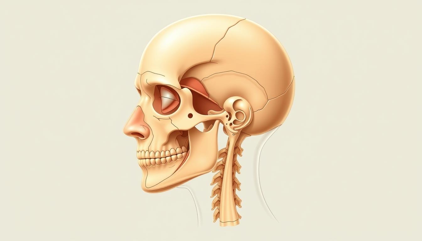
The head’s skeleton includes the cranium and facial bones. Important landmarks are the supraorbital ridge, zygomatic arches, and mandible. These help muscles attach and guide clinical exams.
Soft Tissue Organization
Soft tissues in the head are the skin, muscles, nerves, and blood vessels. Facial muscles, for example, control expressions and get their nerve supply from the facial nerve. How these tissues are organized is important for both normal and abnormal conditions.
“The complex link between the head’s skeletal and soft tissues shows how detailed head anatomy is.”
Functional Divisions in American Medical Practice
In American medicine, the head is split into areas for different specialties. The face, for example, is key for dermatology, plastic surgery, and ear, nose, and throat care. Knowing these areas is essential for diagnosing and treating patients.
Dividing the head into regions helps focus on patient care. The frontal region is often seen in neurosurgery, while the oral region is important for dental and facial surgery.
Head Zone1: Detailed Examination and Clinical Significance
Head Zone1 is a key area that needs careful study for good diagnosis and treatment. It’s important in head diagrams and body maps. It helps us understand the human head’s complexity.
Precise Anatomical Boundaries and Key Structures
The boundaries of Head Zone1 are marked by important landmarks. These landmarks help us know where the zone starts and ends. They also show how it connects with other parts.
- The front boundary is the frontal bone.
- The back boundary is the occipital bone.
- The sides are the temporal bones.
Inside Head Zone1, we find vital parts like the eyes, ears, and blood vessels. These parts are key for our senses and movement.
Neurovascular Supply and Sensory Innervation
The blood and nerve supply to Head Zone1 is complex. It involves arteries, veins, and nerves. The trigeminal nerve is very important for feeling in this area.
- The ophthalmic division of the trigeminal nerve covers the forehead and eyes.
- The maxillary division is for the mid-face.
- The mandibular division is for the lower face and jaw.
Clinical Implications and Common Pathologies
Head Zone1 can face problems like infections, injuries, and brain disorders. Spotting and treating these issues right away is key.
Diagnostic Approaches in U.S. Healthcare Settings
In the U.S., doctors use exams, scans, and tests to diagnose. CT scans and MRI help see injuries and diseases in Head Zone1.
Treatment Considerations
Treatment for Head Zone1 problems depends on the issue. It might need surgery, medicine, or other treatments. A team of experts often works together on tough cases.
Head Zone2: A Detailed Look at Its Medical Importance
Understanding Head Zone2 is key to diagnosing and treating many head-related medical conditions. This area is complex, with many important structures that help our bodies work right.
Precise Anatomical Boundaries and Key Structures
Head Zone2 has clear boundaries that set it apart from other areas. It includes vital structures that are connected and essential for our health.
The edges of Head Zone2 are marked by specific points that can be seen through imaging and studies. Knowing these boundaries is vital for correct diagnosis and treatment.
Neurovascular Supply and Sensory Innervation
The blood vessels and nerves in Head Zone2 form a complex network. This network supplies blood and sensory information. It comes from the external carotid artery and the trigeminal nerve.
This network is key for Head Zone2 to function properly. Any problems with it can cause serious health issues.
Clinical Implications and Common Pathologies
Head Zone2 can be affected by many conditions because of its complex structure and blood supply. These include vascular problems, neurological issues, and injuries.
It’s important to understand these conditions to manage and treat them effectively. The right diagnosis and treatment are essential for Head Zone2 problems.
Diagnostic Approaches in U.S. Healthcare Settings
In the U.S., doctors use several methods to diagnose Head Zone2 issues. These include clinical exams, imaging like MRI and CT scans, and sometimes, more advanced tests. These tools help doctors see the area’s structure and find any problems.
Treatment Considerations
Treatment for Head Zone2 conditions depends on the specific issue and the patient’s health. It might involve medicine, surgery, or other treatments. The choice depends on the diagnosis and the patient’s overall health.
Functional and Clinical Relationships Between Head Zones1 and2
It’s key to know how Head Zones 1 and 2 work together for the right treatment. The brain, hidden inside the skull, is very important in this connection. Using the right words helps us understand how these zones interact.
Anatomical Continuity and Shared Structures
Head Zones 1 and 2 are connected by shared parts. The scalp, for example, covers both zones. The skull’s bones also link them, supporting nerves and blood vessels.
Shared blood supply and venous drainage are key. The external carotid artery feeds the scalp. The internal carotid artery goes deeper, to the brain.
Overlapping Innervation Patterns
The nerves in Head Zones 1 and 2 overlap. The trigeminal nerve plays a big role here. Knowing how these nerves work helps doctors diagnose and treat problems.
Integrated Clinical Assessment Approaches
Assessing Head Zones 1 and 2 together is important. Doctors look at their shared parts, nerves, and how they work together. This helps with treating headaches, facial pain, and other issues.
Using this approach, doctors can make better plans for each patient’s needs.
Head Zone23: In-depth Exploration and Medical Applications
Head Zone23 needs a close look to grasp its medical uses. It has a detailed anatomy that’s key in many medical situations.
Precise Anatomical Boundaries and Key Structures
Head Zone23 has clear front and back limits. The anterior (or ventral) side is the front, and the posterior (or dorsal) side is the back. Knowing these limits helps find important parts in this zone.
This zone holds vital structures like nerves, blood vessels, and landmarks. These are key for its function and head anatomy.
Neurovascular Supply and Sensory Innervation
The nerves and blood vessels in Head Zone23 are complex. They provide sensory info and blood flow. The sensory innervation is vital for sending signals to the brain.
A leading medical expert says, “The complex nerves and blood vessels in Head Zone23 show its role in health and disease” (
This complex interplay between nerves and vessels is key for diagnosis and treatment.
).
Clinical Implications and Common Pathologies
Head Zone23 faces many diseases because of its anatomy and blood supply. These include inflammation, injuries, and neurological issues.
Diagnostic Approaches in U.S. Healthcare Settings
In the U.S., doctors use exams, scans, and tests to diagnose Head Zone23 issues. They follow guidelines to make sure they diagnose and treat correctly.
Treatment Considerations
Treatment for Head Zone23 problems depends on the disease. It might include medicine, surgery, or both. The choice is based on the latest research and the patient’s needs.
Knowing Head Zone23 is essential for anatomy and medicine. Its complex structure and medical importance make it a key area of study in head classification and Head Zones.
Head Zone24: Detailed Analysis and Clinical Relevance
Head Zone24 is complex and needs a detailed look for good care. It’s special because of its detailed structures and big impact on health.
Precise Anatomical Boundaries and Key Structures
Head Zone24 has clear boundaries that are key to understanding its health role. It has important structures that help our body work right.
The edges of Head Zone24 are marked by specific points. These points are vital for finding and treating problems.
Neurovascular Supply and Sensory Innervation
The blood and nerve system in Head Zone24 is complex. Knowing this helps us see how it feels and its health issues.
The nerves in Head Zone24 send out feelings. This is important for its health problems.
Clinical Implications and Common Pathologies
Head Zone24 has many health issues and implications. Knowing these is key for finding and treating problems.
| Pathology | Clinical Implication | Diagnostic Approach |
|---|---|---|
| Trauma | Potential for nerve damage | Imaging studies |
| Infection | Risk of abscess formation | Clinical examination and imaging |
Diagnostic Approaches in U.S. Healthcare Settings
In U.S. healthcare, finding problems in Head Zone24 uses both doctor checks and high-tech scans. These steps are key for right diagnosis and treatment plans.
Treatment Considerations
Treatment for Head Zone24 problems depends on the issue and how bad it is. Options might be watching it, surgery, or both.
Functional and Clinical Relationships Between Head Zones23 and24
It’s important to understand the connection between Head Zones 23 and 24. They have unique features that help the body work right. The body uses membranes and other parts to keep things separate, which is key in the head’s complex structure.
The head is split into different zones, each with its own job. Zones 23 and 24 are special because they work together closely. This teamwork is important for both their structure and how they’re used in medicine.
Anatomical Continuity and Shared Structures
Head Zones 23 and 24 are connected by shared parts. These parts help them work together. They include muscles, blood vessels, and nerves that help the body function.
Shared muscles help move and steady the head. Blood vessels make sure tissues get enough blood. Nerves, both in the head and elsewhere, are key for feeling and moving.
| Structure | Function | Clinical Significance |
|---|---|---|
| Muscular | Movement and stabilization | Injury can lead to impaired mobility |
| Vascular | Blood supply | Compromise can result in ischemia |
| Neural | Sensory and motor innervation | Damage can cause neurological deficits |
Overlapping Innervation Patterns
The nerves that connect Head Zones 23 and 24 overlap a lot. This is key for them to work well together. Both the body’s main nerves and the nerves that control automatic functions are involved.
Knowing about these nerve patterns helps doctors figure out and treat problems. It’s important for them to understand how nerves work together to help patients.
Integrated Clinical Assessment Approaches
Checking Head Zones 23 and 24 together is a smart way to assess them. It looks at their structure, how they work, and any problems they might have. This helps doctors find and fix issues better.
By looking at how these zones are connected, what they share, and their nerve patterns, doctors can make plans to help patients. This way, they can focus on what each patient needs.
The Science of Head Zones Through Body Mapping Techniques
Body mapping has changed how we study head zones. It helps doctors understand the head’s complex anatomy better. By making detailed maps, they can diagnose more accurately and treat patients more effectively.
Principles and Methodology of Body Mapping
Body mapping is about drawing the head’s anatomy with precision. It uses standard terms to avoid confusion. The head’s normal position is the starting point for these detailed drawings.
It involves finding key landmarks and marking the boundaries between different areas. Tools like MRI and CT scans help make these maps more precise.
Creating Comprehensive Body Map Notes for Head Zones
Writing detailed body map notes is key for documenting head zone anatomy. These notes should describe the structures and layers in each area. This way, doctors can keep all important information in one place.
Also, these notes help track changes over time. This is useful for seeing how diseases progress or if treatments are working. It’s very helpful in both treating patients and in research.
Digital Innovations in Body Mapping
New digital technologies have boosted body mapping. Now, software lets us create detailed, interactive 3D models of the head. These models can be rotated and viewed from different sides, giving a deeper look at the head’s anatomy.
Also, digital tools make it easier to share and work on body map data together. This leads to better care coordination and better patient results.
Clinical Applications of Head Zone Knowledge in American Healthcare
Understanding Head Zones is key for better medical care in the U.S. This knowledge helps in diagnosing and treating patients. It’s also important for patient care as children learn about their bodies.
Evidence-Based Diagnostic Considerations
Getting a correct diagnosis is vital in healthcare. Knowing about Head Zones helps doctors do this better. They can spot and treat head and neck problems more accurately.
- Precise identification of anatomical structures
- Understanding of neurovascular supply
- Recognition of clinical implications
Therapeutic Approaches and Intervention Strategies
Dealing with Head Zone issues needs a deep understanding of their anatomy and function. Using proven treatments, doctors can help patients get better and avoid complications.
Key therapeutic considerations include:
- Targeted interventions based on Head Zone anatomy
- Multidisciplinary approaches to patient care
- Monitoring and adjustment of treatment plans
Surgical Planning and Intraoperative Considerations
When it comes to surgeries in the Head Zones, careful planning is essential. Knowing the exact anatomy is key for a successful surgery.
Case Studies from Leading U.S. Medical Centers
Top U.S. medical centers have seen great success in Head Zone surgeries. For example, a leading neurosurgery center showed how knowing anatomy is critical for the best results.
Future Directions in Clinical Practice
The future of Head Zone care will see new imaging tech and more research. As we learn more, we’ll be able to care for patients even better.
Advanced Imaging and Visualization Technologies for Head Zones
Advanced imaging and visualization technologies are key to understanding head zones. Modern medical imaging devices let doctors see “virtual sections” of the body, known as scans. These tools are vital for diagnosing and treating head zone conditions.
Current Radiological Approaches in U.S. Clinical Settings
In the U.S., doctors use different radiological methods to image head zones. Magnetic Resonance Imaging (MRI) and Computed Tomography (CT) scans are top choices. MRI shows soft tissue details, while CT scans are great for bones and calcifications.
These methods have boosted diagnostic accuracy and help plan treatments. For example, MRI is great for looking at the brain’s complex structures. CT scans are often used in emergencies to quickly check for injuries.
3D Modeling and Virtual Reality Applications
3D modeling and virtual reality (VR) have changed how we study head zone anatomy. These technologies create detailed, interactive models. They can be manipulated in real-time, making them useful for both learning and clinical use.
Surgeons use 3D models to plan surgeries. VR offers immersive training for medical students and professionals. This can lead to better patient care by lowering surgery risks.
Integration of Imaging with Body Map Notes
Combining advanced imaging with body map notes has transformed anatomical studies. This method helps doctors understand how different structures in the head zones relate to each other.
This approach improves diagnosis and treatment planning. It also supports research into head zone anatomy, pushing medical science forward.
Interdisciplinary Perspectives on Head Zones in Modern Medicine
Studying Head Zones needs a team effort from many fields, like neurology and neurosurgery. It’s about understanding the brain and its role in our body. This requires a broad view from different medical areas.
Neurology and Neurosurgical Considerations
Neurology and neurosurgery are key in treating Head Zones issues. They focus on the brain, which is hidden inside the skull. Doctors check head functions like feeling and moving.
They might need to operate if there’s a brain injury or tumor. New imaging tools like MRI and CT scans help see the brain better. This makes diagnosing and treating easier.
Otolaryngology and Maxillofacial Surgery Applications
Otolaryngology and maxillofacial surgery deal with ear, nose, throat, and facial issues. They often work with neurology and neurosurgery, mainly for complex injuries.
Understanding the head anatomy is key for these surgeries. Doctors from these fields team up to fix facial fractures, infections, and tumors.
Physical Therapy and Rehabilitation Approaches
Physical therapy and rehab help patients get better after injuries or surgeries. Therapists use special methods to improve head functions like moving and balancing.
Educational Frameworks in U.S. Medical Training
In the U.S., medical schools teach about Head Zones in many subjects, like neurology and surgery. Students learn about head anatomy and its importance in medicine.
| Specialty | Focus Areas | Common Conditions |
|---|---|---|
| Neurology | Diagnosis and treatment of neurological disorders | Stroke, epilepsy, neurodegenerative diseases |
| Otolaryngology | Ear, nose, and throat conditions | Hearing loss, sinusitis, throat infections |
| Maxillofacial Surgery | Surgical interventions for facial and cranial conditions | Facial trauma, oral cancers, congenital defects |
Conclusion
The head is made up of different areas called Head Zones. Each zone has its own features and is important for health. Doctors use special terms, body maps, and new imaging tools to find and fix problems in these areas.
These Head Zones are key in diagnosing and treating many health issues. Knowing how they work together helps doctors find better ways to help patients. This leads to better health results for everyone.
New tools and techniques have changed how we look at head and neck anatomy. They let doctors see the head’s complex parts clearly. This makes it easier to find and treat problems accurately.
To sum up, knowing about Head Zones is vital for doctors to care for patients with head and neck issues. By using what they’ve learned, doctors can do a better job. This means patients get the best care possible.