Learning about cellular organelles is key in biology. It’s important to know the differences between animal and plant cells.
The diagram from ResearchGate shows the Cell Structure of both types. It points out the cell wall in plant cells and its absence in animal cells.
Looking at these differences helps us understand how cells work. This knowledge is vital in biology, medicine, and agriculture.
The Fundamentals of Cellular Biology
Cells are the basic building blocks of life. They are studied in cellular biology. This field looks at how cells are structured, function, and interact.
The Cell as the Basic Unit of Life
The cell is the smallest unit of life. It has key parts like the plasma membrane. This membrane controls what goes in and out. The nucleus holds most of the cell’s genes, guiding its activities.
Cells vary in function but share basic traits. They all have a plasma membrane and need energy to survive.
Historical Development of Cell Theory
The cell theory says all life is made of cells. It was first proposed in the 19th century by Matthias Jakob Schleiden and Theodor Schwann. Their work started modern cellular biology, which now understands cell structures and functions well.
Discovering the cell’s inner parts, like the nucleus, was key. Advances in microscopy helped scientists see cells more clearly. This has greatly helped in understanding how cells work.
Cell Structure: A Detailed Look
Exploring cell structure shows us the complex ways life works at its core. Cells are life’s basic units, and their detailed setup is key to their work.
Eukaryotic vs. Prokaryotic Cell Organization
Eukaryotic cells have a complex setup with a nucleus and organelles like mitochondria. These are the cell’s powerhouses. On the other hand, prokaryotic cells have no nucleus or most organelles, showing a simpler cell form.
Both types of cells have cytoplasm, a jelly-like substance. It’s where many organelles float, helping the cell work.
The Importance of Cellular Compartmentalization
Cellular compartmentalization helps separate different cell processes. This makes things more efficient and reduces confusion between metabolic paths. Organelles like mitochondria and the endoplasmic reticulum are examples of these specialized areas.
Visualizing Cell Structures Through Diagrams
Diagrams are vital for grasping cell structure. They show the complex setup of cell parts. By showing where organelles are in the cytoplasm, diagrams help us understand cell biology better.
ResearchGate’s Contribution to Cell Biology Education
ResearchGate is key in improving cell biology education. It offers a huge collection of scientific diagrams and research. This helps students understand complex biological processes better.
The Role of Scientific Platforms in Knowledge Dissemination
Platforms like ResearchGate are vital for sharing cell biology knowledge. They give researchers and students a lot of information. This includes diagrams of important parts like the endoplasmic reticulum and Golgi apparatus. These sites help people work together and find new things faster.
Accuracy and Detail in ResearchGate Cell Diagrams
The cell diagrams on ResearchGate are very accurate and detailed. They help show how cells work. For example, they show how the endoplasmic reticulum makes proteins and how the Golgi apparatus sorts them. These diagrams are very helpful for both students and researchers.
| Cellular Organelle | Function | Importance in Cell Biology |
|---|---|---|
| Endoplasmic Reticulum | Protein Synthesis and Lipid Metabolism | Critical for cellular homeostasis and function |
| Golgi Apparatus | Protein Modification and Sorting | Essential for cellular secretion and communication |
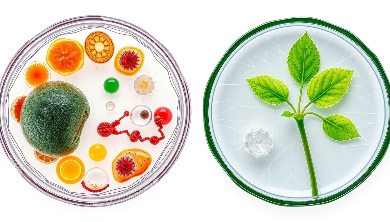
Animal Cell Structure: Defining Characteristics
Animal cells don’t have a cell wall. This makes them very flexible in shape and function. It’s key to understanding their many roles in living things.
Morphological Features of Animal Cells
Animal cells are eukaryotic, meaning they have a nucleus and other organelles. They have a plasma membrane and organelles like lysosomes. These help with digestion and recycling.
These organelles help animal cells stay healthy and react to their environment.
- Presence of membrane-bound organelles
- Absence of a cell wall
- Variable cell shape due to flexibility
Structural Organization and Cellular Architecture
Animal cells have a complex structure. Organelles work together to keep the cell running smoothly. The cytoskeleton supports the cell and helps with division and movement.
The cytoplasm is where many metabolic processes happen. This shows how complex animal cells are.
Specialized Animal Cell Types
Animal cells turn into different types, each with its own job. For example, nerve cells send signals, and muscle cells contract. This specialization is what makes animal cells so important.
- Nerve cells for signal transmission
- Muscle cells for contraction
- Epithelial cells for lining surfaces and glands
Plant Cell Structure: Distinctive Elements
Plant cells are different from animal cells because they have special parts. These parts help them make food through photosynthesis and stay strong. Key features include a tough cell wall, chloroplasts, and a big central vacuole.
Morphological Features of Plant Cells
Plant cells have unique shapes and parts. The cell wall gives them strength and protection. Chloroplasts are key for photosynthesis, letting plants make their own food. The large central vacuole holds water, nutrients, and waste.
- The cell wall is made of cellulose, hemicellulose, and pectin.
- Chloroplasts have chlorophyll, which catches light energy.
- The central vacuole keeps the cell firm and stores stuff.
Structural Organization and Cellular Architecture
Plant cells are organized in a complex way. Organelles work together to keep the cell balanced. The plasma membrane controls what goes in and out. The cytoplasm is where organelles live and move around.
This setup lets plant cells do many important jobs. These jobs help plants grow and develop.
Specialized Plant Cell Types
Plant cells can be different for various tasks. For example:
- Parenchyma cells help with photosynthesis, storing, and making things.
- Collenchyma cells add support and shape.
- Sclerenchyma cells help protect and keep things firm.
These special cells show how diverse and complex plant cells are.
The Plasma Membrane: Cellular Boundary
The plasma membrane is key to a cell’s structure. It acts as a barrier between the cell and the outside world. It helps keep the cell’s contents safe by controlling what goes in and out.
Molecular Composition and Fluid Mosaic Model
The plasma membrane is made mostly of phospholipids and proteins. The fluid mosaic model explains how these molecules are arranged. Phospholipids form a bilayer, with their water-loving heads facing out and their water-fearing tails inside.
This setup makes the membrane semi-permeable. It controls what can pass through, keeping the cell’s contents safe.
Membrane Transport Mechanisms
The plasma membrane manages the flow of substances through different methods. Passive transport like diffusion and osmosis happen without energy. Active transport needs energy to move substances against their natural flow.
The nucleus and cytoplasm are vital in these processes. The nucleus gives the instructions, and the cytoplasm is where most of the action happens.
Comparative Membrane Features in Animal vs. Plant Cells
Animal and plant cells have similar plasma membranes but differ in other ways. Plant cells have a strong cell wall outside the membrane for support. Animal cells, on the other hand, have a more flexible membrane without a cell wall.
Knowing these differences helps us understand the unique traits of each cell type.
The Nucleus: Genetic Control Center
The nucleus is a key part of a cell. It’s like a control center for the cell’s genes. It helps control what the cell does by managing its genes.
Nuclear Envelope and Pores
The nucleus has a double membrane called the nuclear envelope. This envelope has nuclear pores. These pores let materials move in and out, helping the nucleus talk to the rest of the cell.
Chromatin Organization and Nucleolus
Inside the nucleus, chromatin is packed tightly. It turns into chromosomes when the cell divides. The nucleolus is where ribosomes are made. Ribosomes are important for making proteins.
Nuclear Function in Cell Regulation
The nucleus controls how genes are used in the cell. It works with other parts like mitochondria and the endoplasmic reticulum. This helps the cell work right and respond to its environment.
In short, the nucleus is very important for the cell’s genetics and control. It’s a key part of eukaryotic cells.
Cytoplasm: The Living Matrix
Cytoplasm is key for cell life and shape. It’s the area between the cell membrane and the nucleus. It includes the cytosol and many organelles.
The cytosol is the liquid part of cytoplasm. It’s made of water, salts, sugars, and organelles. It helps with chemical reactions and protein making.
Cytosol Composition and Properties
The cytosol is mostly water, 70-90%. This makes it perfect for chemical reactions. It also has ions, sugars, and amino acids for cell work.
| Component | Function |
|---|---|
| Water | Medium for chemical reactions |
| Ions (e.g., K+, Na+, Ca2+) | Regulate various cellular processes |
| Sugars and amino acids | Provide energy and building blocks for proteins |
Cytoskeletal Elements: Microfilaments, Microtubules, and Intermediate Filaments
The cytoskeleton has microfilaments, microtubules, and intermediate filaments. They give structure and help the cell move. Microfilaments help with signals and muscle work. Microtubules keep shape and organize organelles. Intermediate filaments add strength and stability.
Cytoplasmic Streaming in Plant Cells
Cytoplasmic streaming moves cytoplasm in plant cells. It’s driven by the cytoskeleton. This movement spreads organelles and nutrients, helping the cell work well.
In summary, cytoplasm is vital for cell life. It supports many cell functions and keeps the cell balanced. Its complex mix and movement make it a key area in cell biology, linked to the Golgi apparatus and lysosomes.
Mitochondria: Cellular Energy Production
Mitochondria are key in making energy for cells. They are found in most living things, like animals, plants, and fungi.
Structural Features and Compartments
Mitochondria have two main parts: an outer and an inner membrane. The outer membrane lets some things pass through. The inner membrane is folded into cristae, which help make energy. Inside, the matrix holds enzymes for the citric acid cycle.
The Process of Cellular Respiration
Mitochondria make energy for cells through cellular respiration. They break down glucose and other molecules to create ATP. This energy is vital for the cell. The process has three stages: glycolysis, the citric acid cycle, and oxidative phosphorylation.
Mitochondrial Distribution and Abundance in Different Cell Types
How many mitochondria a cell has depends on its energy needs. Muscle cells, needing lots of energy, have more mitochondria. The shape of a cell and where energy is used also affect where mitochondria are found.
Endoplasmic Reticulum: Synthesis and Transport Network
In eukaryotic cells, the cytoplasm is home to the endoplasmic reticulum. This organelle is key for protein synthesis, lipid metabolism, and material transport. It’s a complex structure that supports many cell functions.
Rough Endoplasmic Reticulum: Protein Synthesis
The rough ER has ribosomes on its surface, which are essential for protein synthesis. These proteins are processed and sent to other parts of the cell or outside. It’s mainly found in cells that make a lot of proteins, like pancreatic cells.
Smooth Endoplasmic Reticulum: Lipid Metabolism
The smooth ER doesn’t have ribosomes and focuses on lipid metabolism. It makes cholesterol and phospholipids and detoxifies harmful substances. It also stores calcium ions. Cells in the adrenal gland rely on it for lipid synthesis.
ER-Golgi Transport System
The ER and Golgi apparatus form a transport system vital for cell function. Proteins from the ER go to the Golgi in vesicles. There, they’re modified and sorted for delivery to other parts of the cell or for secretion. This system keeps the cell balanced and ensures proteins and lipids reach their destinations.
| ER Type | Function | Characteristics |
|---|---|---|
| Rough ER | Protein Synthesis | Ribosomes on surface |
| Smooth ER | Lipid Metabolism | No ribosomes |
Golgi Apparatus: Processing and Sorting Center
The Golgi apparatus is a key organelle in eukaryotic cells. It’s in charge of processing proteins and lipids. It makes sure proteins and lipids are modified, sorted, and packaged right for secretion or use inside the cell.
Structural Organization of Cisternae
The Golgi apparatus has a complex structure. It’s made up of flattened sacs called cisternae stacked together. These cisternae are split into three main areas: the cis-Golgi network, the medial-Golgi, and the trans-Golgi network. Each area has its own role in processing proteins and lipids.
Protein Modification and Sorting
The Golgi apparatus is key in modifying proteins and lipids made by the endoplasmic reticulum. It adds carbohydrates to proteins or lipids through glycosylation and phosphate groups through phosphorylation. Then, it sorts these molecules into vesicles for transport to different places in or outside the cell.
Vesicle Formation and Cellular Secretion
The Golgi apparatus packages the modified and sorted molecules into vesicles. These vesicles can merge with the plasma membrane. This releases their contents outside the cell through exocytosis. This process is vital for cell secretion and communication.
| Region | Function |
|---|---|
| Cis-Golgi Network | Receives proteins and lipids from the endoplasmic reticulum |
| Medial-Golgi | Further modification of proteins and lipids |
| Trans-Golgi Network | Sorting and packaging into vesicles for transport |
Lysosomes and Vacuoles: Digestive and Storage Organelles
In the world of cells, lysosomes and vacuoles are key for keeping cells healthy. They help digest and store cell materials. This is vital for cell survival and function.
Lysosomes: Structure and Enzymatic Content
Lysosomes are organelles with digestive enzymes. They break down waste, proteins, lipids, and debris. The acidic inside of lysosomes is needed for their enzymes to work well.
“Lysosomes are the cell’s digestive system,” says Alberts et al., 2002. They are very important for keeping cells in good shape.
Vacuoles: Central Vacuole in Plant Cells vs. Animal Cell Vacuoles
Vacuoles are storage organelles in both plants and animals. But they are different. Plant cells have a big central vacuole for water and salts. This keeps the plant firm.
Animal cells have smaller vacuoles. They help with endocytosis and phagocytosis.
- Plant cell vacuoles are larger and more permanent.
- Animal cell vacuoles are smaller and less permanent.
Autophagic and Digestive Functions
Lysosomes and vacuoles help with autophagy. This is when cells recycle their own parts. Lysosomes break down damaged parts for reuse. This is important for cell health, even when stressed.
As cellular organelles, lysosomes and vacuoles keep the cell clean. They remove waste and recycle nutrients.
Plant-Specific Cellular Components
Plant cells have special parts that help them make food from sunlight. These parts are key for plant cells to live and work well.
Chloroplasts: Structure and Photosynthetic Function
Chloroplasts are where photosynthesis happens in plant cells. They have chlorophyll, which catches sunlight. This sunlight turns carbon dioxide and water into glucose and oxygen.
Chloroplasts have a double membrane and a fluid inside called the stroma. This is where photosynthesis’s light-independent reactions happen.
Cell Wall: Composition and Structural Role
The cell wall is tough and outside the plasma membrane. It supports and protects the plant cell. It’s made mostly of cellulose, which makes it strong.
The cell wall also helps cells talk to each other. It keeps out harmful things too.
| Component | Function |
|---|---|
| Cellulose | Provides strength and rigidity |
| Pectins | Acts as a cementing layer |
| Hemicellulose | Cross-links with cellulose |
Plasmodesmata: Intercellular Communication
Plasmodesmata are tiny channels between plant cells. They let molecules and ions move around. This helps cells talk to each other and work together.
Plasmodesmata are important for sending signals and nutrients between cells. They help plants react to their environment.
In summary, plant cells have special parts like chloroplasts, the cell wall, and plasmodesmata. These parts help plants make food, stay strong, and talk to each other.
Key Differences in Animal vs. Plant Cell Structure
Animal and plant cells have key differences that are important for their roles. Both types of cells have a nucleus and various organelles. But, they have distinct differences in structure and organization.
Morphological and Organizational Distinctions
Plant cells have a cell wall for support and protection. Animal cells do not have a cell wall. Plant cells also have chloroplasts for photosynthesis, while animal cells do not.
Plant cells have large vacuoles for storing water, nutrients, and waste. Animal cells have a flexible cell membrane and centrioles for cilia and flagella. Both types of cells have organelles like mitochondria for energy.
Functional Implications of Structural Differences
The differences in structure affect how animal and plant cells function. Plant cells can make their own food through photosynthesis. Animal cells need to eat other organisms or plants for energy.
The cell wall in plant cells helps maintain their shape and support. The flexible cell membrane in animal cells allows for more movement and flexibility.
| Feature | Animal Cells | Plant Cells |
|---|---|---|
| Cell Wall | Absent | Present |
| Chloroplasts | Absent | Present |
| Large Vacuoles | Absent or small | Present |
Evolutionary Basis for Cellular Divergence
The cell structure differences between animals and plants come from their lifestyles and environments. Plant cells became self-sufficient with photosynthesis. Animal cells became more mobile and responsive.
The cytoplasm is key for both cell types. It helps keep the cell balanced and lets organelles work together.
Modern Techniques for Visualizing Cell Structures
Modern ways to see cell structures have changed how we understand cells. We can now see parts like the endoplasmic reticulum and Golgi apparatus in great detail. This has helped us learn more about how cells work.
Advances in Microscopy
Microscopy has improved a lot, from old light microscopes to new electron microscopes. These new tools let scientists see cells in finer detail. They show us the small parts of cells.
- Light microscopy gives a basic look at cell shapes.
- Electron microscopy shows very detailed images of cell parts.
Fluorescence and Confocal Imaging
Fluorescence and confocal imaging have made it easier to see certain parts of cells. By using special dyes, scientists can watch how these parts move in living cells.
- Fluorescence microscopy lets us see specific cell parts.
- Confocal microscopy gives clear images with less background noise.
3D Reconstruction and Computational Modeling
Using 3D models and computer simulations has helped scientists create detailed cell pictures. These models help predict how cells might act in different situations.
- 3D models help us understand how cells are organized.
- Computer simulations let us predict cell behavior.
Practical Applications of Cell Structure Knowledge
Studying cell structure is key in many fields. Knowing about lysosomes and other cellular organelles helps advance science.
Medical and Pharmaceutical Relevance
In medicine, knowing how cells work is critical. For example, lysosomal dysfunction causes many genetic diseases. Studying cellular organelles can lead to new treatments. Pharmaceutical companies use this info to make drugs that target specific parts of cells.
Agricultural and Biotechnological Implications
In agriculture, understanding plant cells is vital. It helps grow better crops and make plants resistant to pests. Biotech companies use this knowledge to create plants with special traits, like being drought-resistant or more nutritious.
Educational Value of Cell Structure Diagrams
Diagrams of cell structure are great for learning. They help students see how cellular organelles like lysosomes work together. This makes learning about cells easier.
Conclusion
Understanding cell structure is key to knowing how life works at the smallest level. The cell is the basic unit of life. Its parts work together to keep the cell stable and help the organism function.
The plasma membrane is a vital part of cell structure. It controls what goes in and out of the cell. This selective control keeps the cell’s inside environment stable.
In summary, studying cell structure, including the plasma membrane, is very important. It helps us understand how cells work. This knowledge can lead to new treatments for diseases.