The angular incisure is a key spot on the stomach’s lesser curve. It separates the stomach’s body from the pylorus. Knowing about it helps us understand stomach anatomy and how it works.
The angular incisure is linked to the stomach’s structure. It includes pectinate muscles and other muscles. These parts help the stomach digest food.
Looking into the angular incisure and its connections to other parts helps us understand the stomach better. It shows how vital it is in our digestive system.
Anatomical Foundations of the Human Digestive and Cardiovascular Systems
It’s key to know how the body’s digestive and cardiovascular systems work together. These systems are complex and vital for our health. They help keep the body running smoothly.
Integrated Approach to Anatomical Systems
Studying these systems together shows how organs and their functions are connected. The digestive system absorbs nutrients. The cardiovascular system then moves these nutrients around the body.
The muscular components are very important. In the digestive system, muscles help food move. In the cardiovascular system, the heart’s muscles pump blood well.
Muscular Components in Organ Function
The muscular components in the stomach are key for mixing food with digestive enzymes. In the heart, the muscular walls are needed for pumping blood.
| System | Muscular Component | Function |
|---|---|---|
| Digestive | Angular Incisure | Food mixing and churning |
| Cardiovascular | Heart Muscles | Blood circulation |
Knowing how these systems work together is vital. It helps us understand how the body stays healthy and fights off diseases.
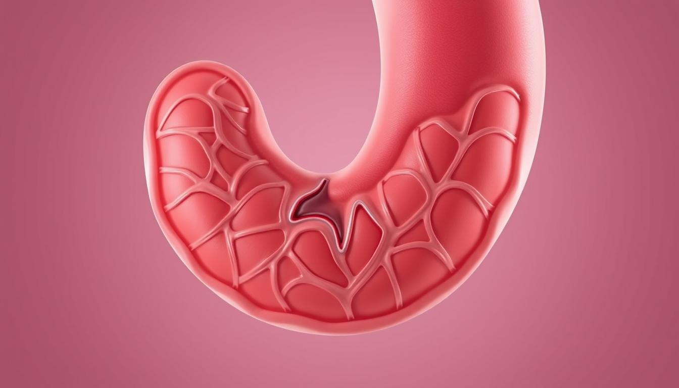
The Angular Incisure of the Stomach: Definition and Location
The angular incisure is found on the lesser curvature of the stomach. It’s a key landmark that helps us understand the stomach’s structure and function.
Anatomical Position and Landmarks
The angular incisure is on the lesser curvature, marking a clear boundary. It’s a key spot for studying anatomy and medicine.
Key aspects of its anatomical position include:
- Location on the lesser curvature
- Serves as a boundary between the gastric body and antrum
- Important for understanding stomach anatomy
Relationship to Gastric Regions
The angular incisure is key in showing where the gastric body meets the antrum. Knowing this helps us grasp the stomach’s anatomy and how it works.
Lesser Curvature Anatomy
The lesser curvature is a major part of the stomach’s anatomy. The angular incisure is a sharp angle that shows where the gastric body and antrum meet.
Transition Between Gastric Body and Antrum
The angular incisure marks the shift from the gastric body to the antrum. This change is vital for understanding the stomach’s function and structure.
The unique anatomy of the angular incisure and its role in the stomach’s regions make it very important.
Histological Features of the Angular Incisure
To understand the angular incisure, we must look closely at its histological details. This part of the stomach is key and has complex features that help it work well.
Tissue Composition and Cellular Organization
The angular incisure has different tissue layers, each with its own cells. The mucosal layer, closest to the inside, has cells that help with digestion and protect against acid.
Muscular Components at the Incisure
The muscles at the angular incisure are mostly smooth muscle.
Smooth Muscle Arrangement
The way smooth muscle is arranged here is important. It helps the stomach move food and mix it with digestive enzymes.
Mucosal and Submucosal Layers
The mucosal and submucosal layers have a special cell and tissue arrangement. The mucosa has gastric glands, and the submucosa has blood vessels and nerves. These are vital for stomach function.
- The mucosa is lined by a simple columnar epithelium.
- Gastric glands in the mucosa produce digestive enzymes and acids.
- The submucosa supports the mucosa with a rich vascular supply.
The histological features of the angular incisure show its role in stomach anatomy and function. Knowing these details helps us understand digestive processes better.
Functional Significance of the Angular Incisure in Digestion
Understanding the angular incisure’s role in digestion is key. This part of the stomach is vital for controlling how fast food moves through. It affects the whole digestive process.
Role in Gastric Emptying and Motility
The angular incisure controls how fast food leaves the stomach. It acts like a gate, letting food into the small intestine at the right pace. This is important for efficient digestion and not overloading the small intestine.
It also impacts stomach movement. The muscles in this area mix food with digestive juices. This helps break down nutrients. The muscles around the angular incisure help create peristaltic waves, moving food through the digestive system.
Peristaltic Wave Formation
Peristaltic waves are vital for food movement. The angular incisure is key in starting and coordinating these waves. The stomach’s muscles, around the angular incisure, create strong contractions. These help mix and move food.
Gastric Content Regulation
The angular incisure also helps manage stomach content. It controls how fast the stomach empties. This prevents too fast or too slow emptying, which can cause problems.
| Function | Description | Impact on Digestion |
|---|---|---|
| Gastric Emptying Regulation | Controls the passage of food into the small intestine | Ensures efficient digestion and prevents overwhelming the small intestine |
| Peristaltic Wave Formation | Initiates and coordinates peristaltic waves | Facilitates the movement of food through the digestive tract |
| Gastric Content Regulation | Regulates the rate of stomach emptying | Prevents rapid or delayed emptying, ensuring optimal digestion |
Muscular Architecture in Hollow Organs: Comparative Analysis
The muscles in hollow organs are designed for their jobs. This part looks at the muscles in the digestive tract and the heart. We’ll see how smooth and striated muscles work together.
Smooth vs. Striated Muscle Distribution
Hollow organs have either smooth or striated muscle. The digestive tract uses smooth muscle for moving food. The heart, with its striated muscle, pumps blood well.
Functional Adaptations of Muscular Tissues
Muscles in hollow organs fit their roles. The digestive tract’s smooth muscle helps food move. The heart’s striated muscle pumps blood all over the body.
Digestive Tract Musculature
The digestive tract has layers of smooth muscle. These layers help mix food and move it along.
Cardiovascular Muscular Components
The heart’s muscles are striated. These cells let the heart beat strongly and work all the time.
| Characteristics | Digestive Tract Musculature | Cardiovascular Musculature |
|---|---|---|
| Muscle Type | Smooth Muscle | Striated Muscle |
| Function | Peristalsis, Mixing Food | Pumping Blood |
| Control | Involuntary | Involuntary |
Looking at the muscles in hollow organs shows how special they are. Knowing these differences helps us understand our bodies better.
Pectinate Muscles: Complete Anatomy and Structure
Pectinate muscles are a key part of the heart’s inside. They are muscular ridges that help the heart work, mainly in the atrial chambers.
Definition and Etymological Origins
The word “pectinate” comes from the Latin “pecten,” meaning “comb.” This is because these muscles look like a comb inside the heart. Their ridged structure is important for their role in the heart’s anatomy.
Structural Characteristics and Arrangement
The way pectinate muscles are arranged is complex and special. They are mostly in the right atrium, adding to its thickness and muscle.
Comb-like Appearance and Organization
The comb-like shape of pectinate muscles helps them contract and relax well. This is key for the heart’s pumping action. Their unique shape is vital for the atrial chambers to work right.
Anatomical Variations
Even though pectinate muscles look the same in most people, they can differ. They can vary in size, number, and where they are in the heart. Knowing these differences is important for studying anatomy and for medical use.
| Characteristics | Description |
|---|---|
| Location | Primarily in the right atrium |
| Structure | Comb-like, ridged |
| Function | Contributes to atrial contraction |
| Variations | Size, number, and location can vary |
In summary, pectinate muscles are a key part of the heart’s anatomy. Their special structure and arrangement are important for the heart’s function. Learning more about them can help us understand heart health and disease better.
Pectinate Muscles in Cardiac Anatomy
The pectinate muscles are key in the heart’s structure, mainly in the atrial chambers. They add to the atrial walls’ complexity and function.
Location Within the Atrial Chambers
Pectinate muscles mostly reside in the right atrium. They can also be found in the left atrium, but less often. Their name comes from their comb-like look, from the Latin “pecten,” meaning comb.
Morphological Features and Development
The pectinate muscles’ shape is important for their role in the heart. They are made of cardiac muscle fibers arranged in a way that helps them contract and relax well.
Relationship to Crista Terminalis
The crista terminalis is a muscular ridge in the right atrium. It separates the smooth-walled back from the rough-walled front, where the pectinate muscles are. These muscles start at the crista terminalis and go into the atrial appendage.
Atrial Appendage Architecture
The atrial appendages, or auricles, are ear-shaped parts of the atria. They have pectinate muscles, which help them contract. This arrangement helps pump blood into the ventricles efficiently.
| Feature | Description |
|---|---|
| Location | Primarily in the right atrium, also in the left atrium |
| Morphology | Comb-like appearance |
| Function | Contributes to atrial contraction and cardiac output |
| Relation to Crista Terminalis | Arise from the crista terminalis |
In conclusion, pectinate muscles are vital in the heart’s anatomy, mainly in the atrial chambers. Their unique structure and location are key to the heart’s function.
Histology and Ultrastructure of Pectinate Muscles
The structure of pectinate muscles is complex, showing their role in heart function. They have a mix of heart muscle cells and connective tissue.
Cellular Components and Organization
Pectinate muscles have heart muscle cells arranged in a special way. These cells are branched and connect to each other. This helps them contract together.
The way these cells are organized is key for the heart’s function. They have lots of myofibrils, which are important for muscle contraction.
Connective Tissue Framework
The connective tissue in pectinate muscles supports their structure. It helps pass on the force of contraction. This tissue is made of collagen and other parts of the extracellular matrix.
Intercalated Discs and Gap Junctions
Intercalated discs are important for linking heart muscle cells. They help spread action potentials and ensure muscles contract together. Gap junctions in these discs let ions and signals move between cells.
Comparison with Ventricular Myocardium
Pectinate muscles and ventricular myocardium share some similarities. But, pectinate muscles have a more complex cell arrangement and a richer connective tissue framework.
| Characteristics | Pectinate Muscles | Ventricular Myocardium |
|---|---|---|
| Cellular Organization | Complex, branched myocytes | More linear, aligned myocytes |
| Connective Tissue | Rich framework | Less prominent |
| Function | Atrial contraction | Ventricular contraction |
Physiological Functions of Pectinate Muscles in Cardiac Cycle
Understanding pectinate muscles is key to knowing their role in heart function and the cardiac cycle. These muscles are unique to the heart, found mainly in the atrial chambers.
Pectinate muscles play a big part in atrial contraction, a key part of the heart’s cycle. They also help with the electrical signals that control heart rhythm.
Role in Cardiac Conduction System
The cardiac conduction system is complex and controls the heartbeat. Pectinate muscles are vital to this system. They help spread electrical signals across the atria.
Contribution to Atrial Contraction
Pectinate muscles boost atrial contraction by adding muscle mass to the atria. This contraction is essential for filling the ventricles with blood before they contract.
Hemodynamic Significance
Pectinate muscles affect atrial pressure and volume. Their role in atrial contraction ensures the ventricles are filled well. This is important for keeping the heart’s output strong.
Electrophysiological Properties
Pectinate muscles have special electrophysiological properties for the cardiac conduction system. Their structure and function help in the efficient spread of electrical impulses. This leads to coordinated atrial contraction.
| Function | Description | Significance |
|---|---|---|
| Cardiac Conduction | Propagation of electrical impulses | Regulation of heart rhythm |
| Atrial Contraction | Enhancement of atrial muscular mass | Optimal ventricular filling |
| Hemodynamic Influence | Impact on atrial pressure and volume | Maintenance of cardiac output |
Clinical Significance of Pectinate Muscles in Cardiovascular Medicine
Pectinate muscles are very important in heart health. They help the heart work right and are key in fighting cardiac arrhythmias and structural heart disease.
Relevance in Cardiac Arrhythmias
Pectinate muscles are linked to heart rhythm problems, like atrial fibrillation. Their complex shape can mess with the heart’s electrical signals.
Atrial Fibrillation Mechanisms
Atrial fibrillation often starts in the pectinate muscles. Knowing how it works helps doctors find better ways to treat it.
Implications in Structural Heart Disease
Pectinate muscles also play a big role in heart structure issues. Problems with these muscles can cause many heart conditions. This makes them important to diagnose and treat.
Considerations in Cardiac Procedures
When doing heart surgeries, knowing about pectinate muscles is key. This knowledge helps doctors work better in complex heart areas. It also helps improve patient results.
A study shows pectinate muscles and heart rhythm issues are closely linked. This calls for better diagnostic tools and treatment plans. It shows the need for more research in heart health and cardiovascular disease.
In summary, pectinate muscles greatly affect heart health. They are vital in managing cardiac arrhythmias and structural heart disease. More research on these muscles is needed to improve heart medicine.
Advanced Imaging Techniques for Studying Muscular Structures
Advanced imaging has changed how we study muscles in the body. These methods let us see and study muscles like the gastric angular incisure and pectinate muscles in detail.
Visualization of Gastric Angular Incisure
The gastric angular incisure is a key area in the body. Advanced imaging helps us see its structure and how it works. This helps doctors diagnose and treat problems related to it.
Cardiac Imaging of Pectinate Muscles
Pectinate muscles are important in the heart. They help the heart work right. We use advanced imaging like echocardiography, MRI, and CT scans to see these muscles.
Echocardiography Applications
Echocardiography is a key tool for checking the heart. It shows the heart in real-time. This lets doctors see how pectinate muscles affect the heart’s performance.
MRI and CT Evaluation Methods
MRI and CT scans give us detailed pictures of the heart. They help doctors see the pectinate muscles and find heart problems. These scans are vital for understanding the heart’s structure.
| Imaging Technique | Application | Benefits |
|---|---|---|
| Echocardiography | Real-time cardiac imaging | Assesses cardiac function and structure |
| MRI | Detailed cardiac anatomy | Evaluates morphology and detects abnormalities |
| CT Scans | High-resolution cardiac imaging | Provides detailed images for diagnosis |
Pathological Conditions Affecting Muscular Components
It’s important to know how diseases affect muscles in organs like the stomach and heart. These muscles are key to how these organs work. When diseases hit these muscles, it can cause a lot of problems.
Disorders of the Angular Incisure
The angular incisure in the stomach can face many issues. Problems like gastric ulcers and cancers can damage it. These issues can start in the lining but spread to the muscle layer.
Gastric Ulcers: Ulcers in the stomach can weaken the angular incisure. This can lead to serious issues like bleeding or holes in the stomach. It can also mess up how the stomach moves food.
Abnormalities of Pectinate Muscles
Pectinate muscles in the heart can also get sick. This includes both genetic problems and diseases that come later in life.
Congenital Variations
Genetic issues with pectinate muscles can change how they work. For example, if there are too many or not enough, it can affect how the heart beats. This might cause irregular heart rhythms.
Acquired Pathologies
Diseases like fibrosis or growth of pectinate muscles can happen later in life. These can be due to heart problems or heart failure. These changes can make heart disease worse.
| Pathological Condition | Affected Structure | Potential Consequences |
|---|---|---|
| Gastric Ulcers | Angular Incisure | Bleeding, Perforation, Impaired Gastric Motility |
| Gastric Cancer | Angular Incisure | Invasion of Muscular Layer, Metastasis |
| Congenital Variations | Pectinate Muscles | Arrhythmias, Altered Atrial Contraction |
| Atrial Fibrillation | Pectinate Muscles | Fibrosis, Hypertrophy, Cardiac Dysfunction |
Understanding these diseases is key to finding better treatments. More research on the muscles in organs and how they react to disease is needed. This will help us manage these conditions better.
Developmental Perspectives: Embryology of Muscular Structures
Understanding how muscles in the stomach and heart develop is key. This knowledge helps us grasp their anatomy and function. The growth of these muscles is complex and vital for the body’s systems to work right.
Formation of Gastric Anatomy
The stomach’s muscles start from the foregut in an embryo. As it grows, the stomach rotates and expands. This creates its unique shape and muscle layers.
The muscularis externa layer forms from the splanchnic mesenchyme. It’s in charge of moving food through the stomach.
Cardiac Chamber Development
Creating the heart’s chambers is a detailed process. It involves many cell types and signals. The heart tube forms early and then loops, separates, and grows into the adult heart.
Molecular signals, like those from the Wnt/β-catenin pathway, guide these steps.
Molecular Signals in Muscle Formation
Molecular signals are vital for muscle growth. For example, the Notch signaling pathway helps decide what cells become in the heart. Growth factors like BMPs and FGFs shape and pattern muscles.
Temporal Sequence of Development
Developmental events must happen in the right order and at the right time. If not, problems can arise. Important steps include:
- The formation of the primitive heart tube
- Looping and septation of the heart
- Differentiation of the muscular layers in the stomach
Recent Research Advances and Future Directions
Research on specialized muscular tissues is changing the game in translational medicine. New studies have greatly improved our understanding of these muscles. This knowledge is leading to new uses in medical science.
Emerging Understanding of Specialized Muscular Tissues
Advanced research is revealing the details of muscular tissues. Specialized muscular tissues are key to many body functions. Knowing how they work is essential for creating better treatments.
Translational Applications in Medicine
Translational medicine is getting a big boost from this research. The insights gained are being used in tissue engineering approaches and therapeutic targeting strategies.
Tissue Engineering Approaches
Tissue engineering is growing fast. It uses knowledge of muscular tissues to create new treatments.
“Tissue engineering has the power to change how we treat muscular disorders. It offers new ways to help patients.”
Therapeutic Targeting Strategies
Therapeutic targeting focuses on finding specific targets in muscular tissues for treatment. This method is leading to treatments that work better and have fewer side effects.
As research keeps moving forward, the future of working with muscular tissues looks bright. It holds promise for many areas of medicine.
Conclusion
The study of the angular incisure and pectinate muscles shows their key role in our body. They are important in the stomach and heart. Knowing about them helps us understand how they work in digestion and heart function.
The angular incisure in the stomach is key for emptying food and moving it. Pectinate muscles in the heart help with heartbeats and how signals move through the heart. These details show how vital they are for our health.
Studying these muscles gives us important information about their function. This knowledge helps us learn more about human anatomy. It also aids in diagnosing and treating related health issues.