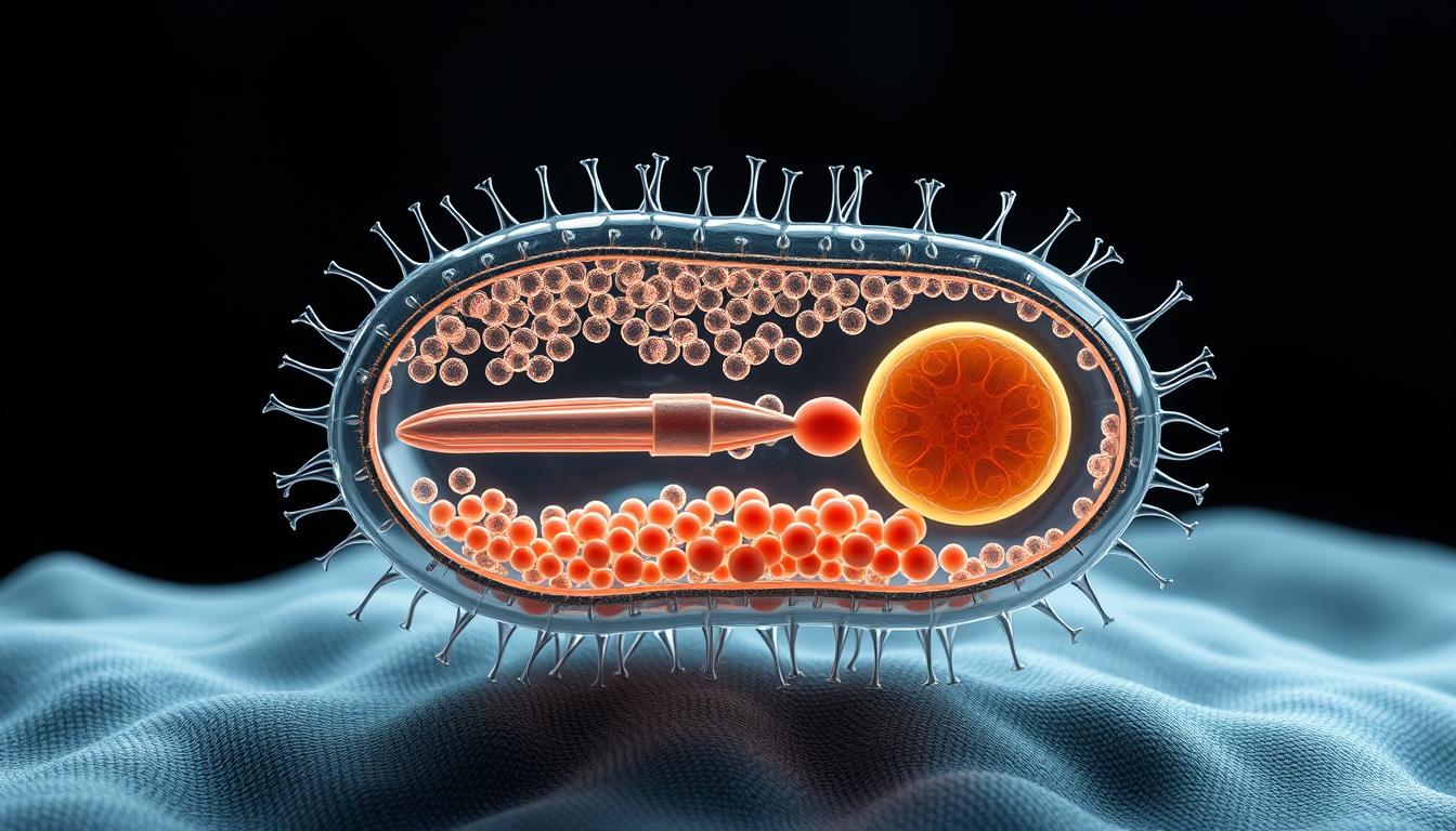
Understanding the prokaryotic cell is key in biology and medicine. The bacterial structure is complex. It has many parts that work together to keep the cell strong.
The bacterial cell diagram shows how it’s organized. It helps researchers and students understand its functions and how it interacts. This article will explain the parts of the bacterial cell, as shown in its diagram.
By looking at the bacterial cell structure, we learn about its behavior and how it reacts to different things. This helps us understand these tiny organisms better.
The Bacterial Cell: Fundamental Characteristics
To understand bacterial cells, we must explore their key traits. They lack a true nucleus and other organelles, unlike eukaryotic cells. This prokaryotic nature makes their structure simple but their function complex.
Prokaryotic Nature and Basic Features
Bacterial cells mainly have a cell envelope. This includes the bacterial cell wall and bacterial cell membrane. The cell wall gives support, and the membrane controls what enters and leaves the cell. Without a nucleus, their DNA is in a single circular chromosome in the nucleoid region.
Inside, there’s a cytoplasm where metabolism happens. They also have inclusion bodies for storing nutrients or doing specific tasks.
Size, Shape, and Diversity
Bacteria come in all sizes and shapes, showing their incredible variety. They can be round (cocci), long and thin (bacilli), or spiral (spirochetes). Their shape often matches their environment and lifestyle.
| Shape | Description | Examples |
|---|---|---|
| Cocci | Spherical or oval | Staphylococcus aureus |
| Bacilli | Rod-shaped | Escherichia coli |
| Spirochetes | Spiral-shaped | Treponema pallidum |
Evolution and Classification of Bacteria
Bacteria have been around for over 3.5 billion years. They play a key role in our planet’s ecosystems. Their ability to adapt and diversify is impressive.
Evolutionary History of Prokaryotes
The story of prokaryotes’ evolution is complex. Studies show bacteria were among the first life forms. They can survive in many environments, leading to diverse metabolic processes.
Bacterial DNA replication is vital for their genetic survival. This process is tightly controlled. It’s a key part of how bacteria divide.
Major Taxonomic Groups and Classification Systems
Bacteria are sorted into groups based on their traits. These include cell wall type, metabolic processes, and genetics. The Gram staining method is a common way to classify them.
| Characteristic | Gram-Positive Bacteria | Gram-Negative Bacteria |
|---|---|---|
| Cell Wall Composition | Thick peptidoglycan layer | Thin peptidoglycan layer, outer lipid membrane |
| Gram Staining | Retains crystal violet stain | Does not retain crystal violet stain |
| Examples | Staphylococcus aureus, Bacillus subtilis | Escherichia coli, Pseudomonas aeruginosa |
Understanding bacteria’s classification is important. It helps us see their roles in ecosystems and how they interact with others. Bacterial division is essential for their growth and adaptation to new environments.
Reading a Bacterial Cell Schematic Diagram
To understand bacterial cell biology, you need to read schematic diagrams well. These diagrams are key to grasping how bacterial cells work. They show the details of structures like the cell envelope, bacterial cytoplasm, and organelles.
Common Symbols and Conventions
Bacterial cell diagrams use standard symbols for different parts. For example, the cell wall is shown as a thick layer around the cell membrane. Knowing these symbols is vital for correct interpretation. Bacterial ribosomes are usually small dots in the cytoplasm, showing their role in making proteins.
Diagrams also show important structures like flagella, pili, and the nucleoid. Knowing these symbols helps researchers and students quickly understand bacterial cell structure and function.
Interpreting Cross-Sectional Views
Cross-sectional views in diagrams give a detailed look at the cell’s inside. These views are key to seeing how different parts are arranged. For instance, they might show the cell wall’s thickness or where bacterial ribosomes are in the cytoplasm.
Understanding these views means knowing the cell’s layout and how to see 3D structures from 2D diagrams. By getting good at reading cross-sections, you can dive deeper into bacterial cell complexity.
The Bacterial Cell Envelope
Understanding the bacterial cell envelope is key to knowing how bacteria work and cause disease. The cell envelope is a complex structure that protects the bacterial cell. It also controls what materials move in and out of the cell.
Outer Membrane in Gram-Negative Bacteria
The outer membrane is special in Gram-negative bacteria. It adds extra protection against stress and antibiotics. It’s made of lipopolysaccharides (LPS) and proteins.
Lipopolysaccharides (LPS)
LPS, or endotoxin, is a key part of the outer membrane. It causes strong immune reactions in hosts. It has a lipid A part, a core polysaccharide, and an O-antigen chain.
Porins and Transport Proteins
Porins are proteins that make channels in the outer membrane. They let molecules pass into the periplasmic space. Other transport proteins help specific substrates move across the membrane.
Periplasmic Space and Its Functions
The periplasmic space is between the inner cytoplasmic membrane and the outer membrane in Gram-negative bacteria. It has enzymes and proteins for nutrient processing, cell wall synthesis, and stress response.
| Function | Description |
|---|---|
| Nutrient Processing | Enzymes in the periplasmic space break down complex nutrients into simpler molecules. |
| Cell Wall Synthesis | Proteins involved in peptidoglycan synthesis are located in the periplasmic space. |
| Stress Response | The periplasmic space contains proteins that help bacteria respond to environmental stresses. |
Capsules and Slime Layers
Capsules and slime layers are outer layers made of polysaccharides. They surround many bacterial species. They are important for bacterial virulence, protecting bacteria from drying out, being eaten by phagocytes, and environmental stresses.
The bacterial cell envelope is vital for bacterial survival and causing disease. Knowing about its parts and functions is key to fighting bacterial infections.
Bacterial Cell Wall Structure and Function
Understanding the bacterial cell wall is key to knowing how bacteria work. The cell wall gives mechanical support, keeps the cell’s shape, and stops it from bursting due to pressure.
The cell wall is vital for bacteria to survive. Its makeup changes a lot between different bacteria. A big part of the cell wall is peptidoglycan, made of sugars and peptides.
Peptidoglycan Composition and Assembly
Peptidoglycan is made of N-acetylglucosamine (NAG) and N-acetylmuramic acid (NAM) units. These units are linked by peptide chains. The making of peptidoglycan is important for the cell wall’s strength.
| Component | Function |
|---|---|
| N-acetylglucosamine (NAG) | Provides structural framework |
| N-acetylmuramic acid (NAM) | Linked to peptide chains for cross-linking |
| Penicillin-binding proteins (PBPs) | Catalyze cross-linking of peptidoglycan |
Gram-Positive vs. Gram-Negative Cell Walls
Gram-positive and Gram-negative bacteria have different cell walls. Gram-positive bacteria have a thick peptidoglycan layer that keeps the stain used in Gram staining. Gram-negative bacteria have a thinner layer and an outer membrane with lipopolysaccharides.
“The difference in cell wall structure between Gram-positive and Gram-negative bacteria is not just a matter of thickness; it fundamentally affects their interaction with the environment and their susceptibility to antibiotics.”
Gram-positive bacteria, like Staphylococcus aureus, have a thick layer for support. Gram-negative bacteria, such as Escherichia coli, have a more complex envelope. This envelope includes porins and lipopolysaccharides, helping them resist antibiotics.
Cell Wall Synthesis and Antibiotic Targets
Building the cell wall is key for bacterial growth and division. Antibiotics like penicillin and vancomycin stop peptidoglycan synthesis. This is important for finding new ways to fight bacteria.
Antibiotic resistance, like beta-lactamases breaking down penicillin, shows we need more research. We must keep studying bacterial cell walls and finding new antibiotics.
The Bacterial Cell Membrane
The bacterial cell membrane is vital for keeping the cell together and working right. It’s a complex part that lets substances in and out. This is key for the cell to survive.
Phospholipid Bilayer Architecture
The cell membrane is mainly made of a phospholipid bilayer. The hydrophilic heads are on the outside, and the hydrophobic tails are on the inside. This setup makes a barrier that lets certain molecules pass through.
“The phospholipid bilayer is a critical component of the bacterial cell membrane, providing structural integrity and fluidity,” as noted in scientific literature.
Membrane Proteins and Their Functions
Membrane proteins are in the phospholipid bilayer and do many jobs. They help with transport, signaling, and how cells talk to each other. These proteins are key for the cell to interact with its surroundings.
Transporters and Channels
Transporters and channels are proteins that help substances move across the membrane. They let the cell get what it needs and get rid of waste.
Receptors and Signal Transduction
Receptors on the membrane bind to specific molecules, starting signal pathways. This helps the cell respond to changes in its environment. It’s important for the cell to adapt.
Membrane Potentials and Energy Generation
The membrane is also important for making energy. It creates a membrane voltage that helps with energy production. This voltage is essential for many cell functions, like making ATP.
The membrane voltage comes from ions moving across the membrane. This creates an electrochemical gradient. The cell uses this gradient to make energy.
Bacterial Cytoplasm and Its Contents
The cytoplasm of a bacterial cell is a busy and organized space. It’s where important activities happen, like making proteins and storing nutrients. This complex environment is key for the cell’s survival and function.
Cytosol Composition and Properties
The cytosol is the liquid part of the cytoplasm, taking up a lot of the cell’s space. It’s like a gel made of water, salts, sugars, and organic compounds. This liquid helps enzymes work, making the cell run smoothly.
Inclusion Bodies and Storage Granules
Inclusion bodies and storage granules are special parts in the cytoplasm. They store nutrients and waste. These structures help the cell survive in different conditions.
Polyphosphate Granules
Polyphosphate granules store phosphate, a vital nutrient for bacteria. They’re very important when phosphate is scarce.
Glycogen and Other Storage Compounds
Glycogen is a complex sugar that acts as energy storage in some bacteria. Other compounds, like sulfur granules, may also be present, depending on the bacteria and its environment.
Gas Vesicles
Gas vesicles are found in some aquatic bacteria. They’re made of protein and help the bacteria stay afloat in water.
Bacterial Cytoskeleton
The bacterial cytoskeleton is a network of proteins that gives the cell shape and support. It also helps with cell division and moving genetic material.
In summary, the bacterial cytoplasm is a dynamic and organized space. It’s vital for the cell’s survival and function. The cytosol, inclusion bodies, and cytoskeleton work together to support metabolic processes, store nutrients, and keep the cell balanced.
The Bacterial Nucleoid and Genetic Material
Bacteria have a unique nucleoid region for their genetic material. This is key to understanding how they adapt and evolve.
Bacterial Chromosome Organization
The bacterial chromosome is a single, circular DNA molecule. It’s found in the nucleoid region. This DNA is compacted through supercoiling and nucleoid-associated proteins (NAPs).
The chromosome’s organization is vital for DNA replication and gene expression. Unlike eukaryotic cells, it’s not membrane-bound. This allows for direct interaction between DNA and the cytoplasm.
NAPs are important for the nucleoid structure. They help compact DNA and affect gene expression. The chromosome’s organization can change between species and growth phases.
DNA Replication in Bacterial Cells
DNA replication is essential for bacterial cells. It ensures each daughter cell gets a complete genome. The process starts at a specific origin and moves in both directions.
Many proteins work together to replicate DNA. The cell makes sure replication is done before dividing. This keeps the genome intact. Bacterial DNA replication is also a target for antibiotics, showing its importance.
Plasmids and Mobile Genetic Elements
Bacteria often have plasmids, smaller DNA molecules. Plasmids carry genes for traits like antibiotic resistance. They can move between bacteria, spreading genetic information.
Mobile genetic elements, like transposons and integrons, are also important. They can move within or between genomes. This can change gene function or introduce new traits, adding to bacterial diversity.
Bacterial Ribosomes and Protein Synthesis
Ribosomes are key in making proteins in bacteria. They are complex machines that turn mRNA into amino acids. These amino acids then form proteins.
Structure of 70S Ribosomes
Bacterial ribosomes are called 70S ribosomes. They have two parts: the 50S and 30S subunits. The 50S part does the peptidyl transferase activity. The 30S part decodes the mRNA.
The 70S ribosomes are made mostly of ribosomal RNA (rRNA) and proteins. The rRNA gives the structure. The proteins help stabilize and facilitate the translation process.
Translation Process in Prokaryotes
Translation in bacteria starts with the mRNA binding to the 30S subunit. Then, the 50S subunit joins. The process adds amino acids to a polypeptide chain. It ends when a stop codon is found.
“The ribosome is a molecular machine that reads the sequence of messenger RNA and translates it into a specific sequence of amino acids, building proteins.”
The translation process in bacteria is different from eukaryotes. This difference is why some antibiotics target bacterial ribosomes.
Ribosomal Antibiotics and Their Mechanisms
Many antibiotics work by stopping protein synthesis in bacteria. Tetracyclines, aminoglycosides, and macrolides are examples. Tetracyclines stop aminoacyl-tRNA from binding. Aminoglycosides cause mistakes in amino acid incorporation. Macrolides block the exit tunnel for the polypeptide chain.
| Antibiotic Class | Ribosomal Target | Mechanism of Action |
|---|---|---|
| Tetracyclines | 30S subunit | Prevents aminoacyl-tRNA binding |
| Aminoglycosides | 30S subunit | Causes misreading of mRNA |
| Macrolides | 50S subunit | Blocks exit tunnel for polypeptide chain |
These antibiotics target bacterial ribosomes because of their differences from eukaryotic ribosomes. This allows for effective treatment with less harm to host cells.
Bacterial Cell Surface Appendages
Surface appendages on bacterial cells are vital. They help with motility, adhesion, and environmental interactions. These structures are key for bacteria to survive and thrive.
Flagella Structure and Bacterial Motility
Flagella are complex, whip-like structures. They enable bacteria to move through their environment. The structure and function of flagella are critical for bacterial motility.
Flagellar Assembly
The assembly of flagella involves the coordinated synthesis and transport of various proteins. This process is highly regulated and essential for the proper functioning of flagella.
Chemotaxis and Directed Movement
Chemotaxis is the ability of bacteria to move towards or away from certain chemicals in their environment. This directed movement is key for finding nutrients and avoiding harmful substances.
Pili and Fimbriae: Structure and Functions
Pili and fimbriae are hair-like appendages on the surface of bacterial cells. They play significant roles in adhesion, DNA transfer, and other cellular processes.
Surface Layer Proteins (S-layer)
The S-layer is a crystalline layer composed of proteins or glycoproteins that covers the surface of some bacteria. It provides structural support and protection, and is involved in various interactions with the environment.
In conclusion, bacterial cell surface appendages are essential for various aspects of bacterial behavior and survival. Understanding these structures is key to comprehending bacterial interactions with their environment.
Bacterial Cell Division and Reproduction
Bacteria reproduce through binary fission. This method ensures their genetic continuity. It allows them to grow quickly when conditions are right.
Binary Fission Process
Binary fission starts with the replication of the bacterial chromosome. After DNA is copied, the cell gets ready to divide. It elongates and forms a septum at the middle.
The septum is key to separating the two new cells. Proteins and enzymes work together in this process. For example, DNA replication begins at a specific point called the origin of replication.
As division approaches, the chromosomes move to opposite ends of the cell.
FtsZ Ring and Division Machinery
The FtsZ ring is vital for division. It’s made of FtsZ proteins and forms at the division site. It helps the cell membrane to narrow and the septum to form.
Proteins like FtsA and ZipA help keep the FtsZ ring stable. They also bring in more division proteins. Together, they form the divisome, which drives division.
Regulation of Cell Division Cycle
The cell division cycle in bacteria is carefully managed. It ensures DNA replication and cell division happen together. Checkpoints in the cycle watch for important steps like DNA replication and chromosome segregation.
Regulators like the Min proteins and SlmA control the FtsZ ring’s formation and placement. They help make sure division is accurate and the new cells are genetically the same.
In summary, bacterial cell division is a complex and regulated process. It’s essential for bacteria to reproduce and grow. Studying binary fission helps us understand bacteria better and find new treatments.
Specialized Bacterial Structures and Adaptations
Bacteria have special structures that help them live in different places. These features let them survive and grow in many environments. This includes places with extreme temperatures and high salt levels.
Endospores and Sporulation Process
Some bacteria make endospores to survive harsh conditions. Endospores are very tough and can handle high temperatures, radiation, and chemicals. The process of making endospores, called sporulation, is complex.
When it’s hard for bacteria to find food, they start making endospores. This process changes the bacterial cell a lot. It includes making a spore septum and adding protective layers around the spore.
Magnetosomes and Bacterial Magnetotaxis
Some bacteria have magnetosomes, which are special parts that help them move. Magnetosomes contain magnetic minerals. This lets bacteria move along magnetic fields.
Magnetotaxis helps bacteria find their way, mainly in water where oxygen levels change. Moving along magnetic fields helps them reach better places.
| Structure | Function | Example |
|---|---|---|
| Endospores | Survival under extreme conditions | Bacillus subtilis |
| Magnetosomes | Magnetotaxis, navigation | Magnetospirillum gryphiswaldense |
| Unique membrane structures | Survival in extreme environments | Thermus thermophilus |
Unique Structures in Extremophiles
Extremophilic bacteria have special features for living in extreme places. For example, thermophilic bacteria have special lipids in their membranes. These lipids keep their membranes stable at high temperatures.
Other extremophiles, like halophilic bacteria, live in very salty places. They have special ways to handle salt and keep their cells balanced.
These special structures and ways of adapting show how diverse and strong bacterial life is. They can live almost anywhere on Earth.
Conclusion: The Remarkable Complexity of Bacterial Cells
The bacterial cell is truly fascinating. It shows a remarkable complexity and diversity. This is key for its survival and function. It has structures like the cell envelope, cytoplasm, and genetic material.
These structures help the cell carry out important tasks. Tasks like metabolism, DNA replication, and protein synthesis are done here. This shows how complex and sophisticated these cells are.
Understanding the bacterial cell’s structure is vital. It helps us see how these microorganisms work. Knowing this is important for fighting bacterial infections.
Studying the bacterial cell is essential in microbiology. It gives us insights into these organisms and their environment. By understanding the bacterial cell, researchers can find new ways to help and innovate.