Diagnosing genetic syndromes needs a deep understanding of their signs. Noonan syndrome is one such condition. It has unique facial features and physical anomalies.
Getting the diagnosis right is key for proper care. Clinical images are very important. They help doctors spot the signs of Noonan syndrome and similar conditions.
This article will look at how clinical images help in diagnosing genetic syndromes, focusing on Noonan syndrome. By studying these images, doctors can better understand the condition. This helps improve patient care.
Understanding Noonan Syndrome and Its Clinical Spectrum
Noonan syndrome is an autosomal dominant disorder. It’s tricky to diagnose because it affects people differently. People with this condition often have facial dysmorphism, heart problems, and may develop slowly.
Historical Perspective and Discovery
Noonan syndrome was first noted in the 1960s. Back then, doctors saw unique physical traits and heart issues. Today, we know more about it, thanks to both medical and genetic research.
Prevalence and Epidemiology
It’s thought that about 1 in 1,000 to 1 in 2,500 babies are born with Noonan syndrome. But, it might be more common than we think. Studies show it affects both boys and girls equally, with no bias towards any race. It can lead to heart problems and developmental delays.
Knowing about the clinical spectrum of Noonan syndrome is key. Each person with it is different, so a detailed check-up is needed.
Genetic Basis of Noonan-like Features
Noonan-like features come from mutations in the RAS/MAPK signaling pathway. This pathway is key for cell growth, development, and survival. When genes in this pathway mutate, it can cause Noonan syndrome and similar conditions.
RAS/MAPK Pathway Mutations
Mutations in the RAS/MAPK pathway are linked to Noonan syndrome and other RASopathies. The main genes affected are PTPN11, SOS1, RAF1, and KRAS. These changes can either activate or block the pathway, messing up cell function.
| Gene | Function | Mutation Effect |
|---|---|---|
| PTPN11 | Encodes SHP2 protein | Activating mutation |
| SOS1 | Encodes SOS1 protein, a RAS guanine nucleotide exchange factor | Activating mutation |
| RAF1 | Encodes RAF1 protein, a serine/threonine kinase | Activating mutation |
Genetic Heterogeneity in Noonan-like Syndromes
Noonan-like syndromes have many genes and mutations involved. This makes diagnosis and genetic counseling tricky. Knowing the exact genetic cause is key for managing the condition and understanding the risk of passing it on.
Genotype-Phenotype Correlations
Genotype-phenotype correlations in Noonan-like syndromes are complex. Some mutations are linked to specific symptoms, like heart problems or developmental delays. Figuring out these connections helps predict the condition’s course and plan treatment.
In summary, Noonan-like features stem from RAS/MAPK pathway mutations, with a lot of genetic variation. Grasping these genetic roots is vital for diagnosis, treatment, and genetic counseling.
Diagnostic Criteria for Noonan-like Features
Diagnosing Noonan syndrome is complex. It involves looking for specific facial features, developmental delays, and other signs. A detailed clinical check and genetic tests are needed to confirm the presence of certain gene mutations.
Van der Burgt Criteria
The Van der Burgt criteria are key for diagnosing Noonan syndrome. They look for facial dysmorphia, heart problems, developmental delays, and other signs. Features like pulmonary stenosis and hypertrophic cardiomyopathy are very important.
Age-Related Manifestations
Noonan syndrome shows different signs at different ages. Babies often have trouble feeding and don’t grow well. Kids may have delays in development and learning. Adults might face heart problems and a higher risk of cancer.
| Age Group | Common Manifestations |
|---|---|
| Infancy | Feeding difficulties, failure to thrive |
| Childhood | Developmental delays, learning disabilities |
| Adulthood | Cardiovascular complications, increased cancer risk |
Variability in Clinical Expression
Noonan syndrome can show up differently in people, even in the same family. This is because of the genetic variety and the effect of other genes. It’s important to understand this to make an accurate diagnosis and care plan.
In summary, diagnosing Noonan syndrome is a detailed process. It includes clinical checks, genetic tests, and knowing about the syndrome’s variability. The Van der Burgt criteria help in diagnosis. Being aware of how the syndrome changes with age and its variability ensures the best care for those affected.
Characteristic Craniofacial Dysmorphology
Noonan syndrome is known for its unique craniofacial features. These features are key in diagnosing the condition. They can be spotted through a detailed clinical check-up.
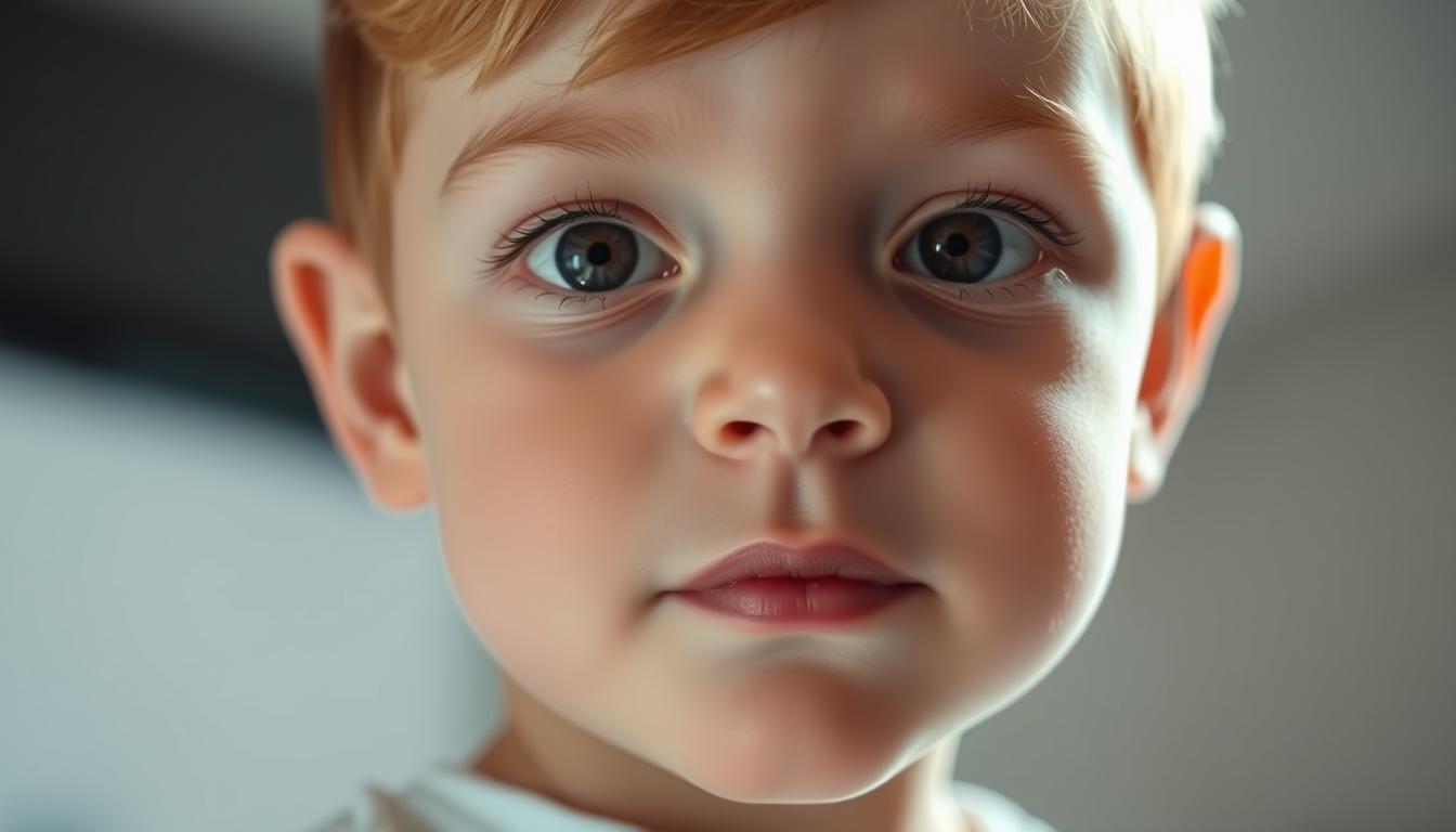
Facial Features Through Clinical Photography
Clinical photos are vital in capturing the faces of those with Noonan syndrome. Characteristic facial features include a wide forehead, eyes that are too far apart, and a broad nose. These traits are more noticeable in kids and can change as they grow older.
Using the same photography methods helps compare faces at different ages. This makes it easier to track changes over time.
Ocular Abnormalities and Imaging
Ocular issues are common in Noonan syndrome. Hypertelorism and ptosis are seen often. Imaging, like orbital scans, helps measure how much these issues affect the eyes.
| Ocular Abnormality | Frequency | Clinical Significance |
|---|---|---|
| Hypertelorism | Common | Affects facial appearance |
| Ptosis | Frequent | May require surgical intervention |
Dental and Oral Manifestations
Dental and oral signs are also important in Noonan syndrome. High-arched palate and dental malocclusion are common. Regular dental visits are key to managing these problems.
The facial, ocular, and oral traits of Noonan syndrome are complex. A thorough clinical evaluation, including photos and scans, is needed for a correct diagnosis and treatment plan.
Cardiovascular Manifestations and Imaging Findings
The heart is often affected in people with Noonan syndrome. This leads to various health issues. It’s important to evaluate and manage these problems well.
Echocardiographic Features of Pulmonary Stenosis
Pulmonary stenosis is common in Noonan syndrome. Echocardiography helps diagnose and measure its severity. Key features include:
- Valvular pulmonary stenosis with characteristic doming
- Variable degrees of stenosis, from mild to severe
- Potential for associated right ventricular hypertrophy
Imaging of Hypertrophic Cardiomyopathy
Hypertrophic cardiomyopathy (HCM) is a big issue in Noonan syndrome. Echocardiography and cardiac MRI are key for diagnosing and tracking HCM.
| Imaging Modality | Key Features in HCM |
|---|---|
| Echocardiography | Asymmetric septal hypertrophy, left ventricular outflow tract obstruction |
| Cardiac MRI | Detailed assessment of hypertrophy, fibrosis, and outflow tract obstruction |
Other Cardiac Anomalies on Imaging
Other heart problems can also occur in Noonan syndrome. These include atrial septal defects and ventricular septal defects. Detailed imaging is key to spotting these issues and helping with treatment.
In summary, heart problems are a big part of Noonan syndrome. They need careful checking and treatment. Advanced imaging is very important for diagnosing and managing these heart issues.
Skeletal and Growth Abnormalities: Radiographic Features
Radiographic analysis is key to understanding skeletal and growth issues in Noonan-like syndrome. These issues can greatly affect a person’s quality of life. So, early diagnosis and treatment are very important.
Growth Charts and Patterns
Growth charts are useful for tracking growth in people with Noonan-like syndrome. They help doctors see if a person’s height, weight, and other growth measures are normal. People with Noonan syndrome often grow shorter, and growth charts help manage this.
A study in the Journal of Medical Genetics showed how important it is to watch growth. It found that growth charts can help decide if someone needs growth hormone therapy.
“Growth hormone treatment in Noonan syndrome: a systematic review,” Journal of Medical Genetics, 2020
Chest Wall Deformities on Imaging
Chest wall issues like pectus excavatum or carinatum are common in Noonan-like syndrome. X-rays and CT scans are used to see how severe these problems are. This helps doctors plan for surgery if needed.
Spine and Extremity Abnormalities
People with Noonan-like syndrome may have spine and limb problems, like scoliosis and joint hypermobility. X-rays and other scans are key for diagnosing and tracking these issues. Below is a table of common radiographic findings in Noonan-like syndrome.
| Abnormality | Radiographic Finding |
|---|---|
| Scoliosis | Curvature of the spine on X-ray |
| Pectus Excavatum | Depression of the sternum on CT scan |
| Joint Hypermobility | Increased joint laxity on stress X-rays |
Knowing these radiographic features is vital for managing Noonan-like syndrome. It helps doctors provide better care and improve patient outcomes.
Clinical Photography of Noonan-like Features in Patients
Clinical photography is key in diagnosing and managing patients with Noonan-like features. It helps doctors document the facial dysmorphism and other physical traits linked to this genetic syndrome.
Standardized Photography Techniques
Using standardized photography techniques is vital for consistency. This means using a top-notch camera, the right lighting, and a steady background. Standardization is important for tracking changes and comparing features among patients.
Key Features to Document
When taking photos of patients with Noonan-like features, focus on specific facial traits. These include the shape of the eyes, the nose’s structure, and the face’s overall proportions. Below is a table outlining these features and their importance.
| Feature | Description | Significance |
|---|---|---|
| Eye Shape | Wide-set eyes | Common in Noonan-like syndrome |
| Nasal Structure | Depressed nasal bridge | Characteristic feature |
| Facial Proportions | Long philtrum | Associated with genetic syndrome |
Evolution of Features with Age
Noonan-like features can change a lot as a patient gets older. Taking photos at different times can show how these changes happen. For example, some facial features might grow more noticeable or change in shape over time.
By capturing these changes, doctors can get a clearer picture of Noonan-like syndrome’s natural progression. This helps them adjust treatment plans more effectively.
Neurological and Developmental Imaging
Neurological and developmental imaging are key in checking for delays in patients with Noonan syndrome. MRI is a top tool for spotting and handling the brain issues linked to the syndrome.
Brain MRI Findings
Brain MRI is a vital tool for finding brain problems in Noonan syndrome. Common issues include:
- Ventricular dilatation: Big ventricles in the brain.
- Cortical dysplasia: Issues with the brain’s outer layer.
- White matter changes: Problems with the brain’s white matter.
These findings help doctors understand the brain effects of Noonan syndrome.
Neuropsychological Assessment Correlation
Linking brain MRI results with neuropsychological tests is key. It helps in:
- Finding out what a person can do well and what they struggle with.
- Creating plans for therapy that really work.
- Improving care for patients.
Motor Development Documentation
Tracking how well a person moves is important in managing Noonan syndrome. Imaging and tests help see how motor skills grow. They also show where extra help might be needed.
Using imaging and tests in treating Noonan syndrome helps doctors understand it better. This leads to better ways to manage the condition.
Hematological and Lymphatic System Abnormalities
Noonan syndrome is an inherited condition that affects the blood and lymphatic systems. These issues can greatly impact a person’s life. They need careful management to improve their quality of life.
Laboratory Findings in Bleeding Disorders
Bleeding disorders are common in Noonan syndrome. They require detailed lab tests to diagnose. Key findings include:
- Coagulation factor deficiencies
- Platelet dysfunction
- Prolonged bleeding times
These lab results are essential for treating bleeding disorders in Noonan syndrome patients.
Imaging of Lymphatic Dysplasias
Lymphatic dysplasias are a big part of Noonan syndrome. Ultrasound and MRI are key for diagnosing and understanding these issues.
Good imaging helps plan the right treatments. It also helps manage problems related to lymphatic dysplasias.
Differential Diagnosis of RASopathies with Noonan-like Features
Understanding RASopathies is key to diagnosing Noonan-like syndrome correctly. RASopathies are genetic disorders linked to the RAS/mitogen-activated protein kinase (MAPK) pathway. This pathway is vital for cell growth, division, and survival.
When diagnosing RASopathies with Noonan-like features, several genetic syndromes come into play. These syndromes share traits like facial features, heart issues, and developmental delays.
Cardio-facio-cutaneous Syndrome
Cardio-facio-cutaneous syndrome is a RASopathy with heart problems, unique facial traits, and skin issues. It’s caused by mutations in the BRAF, MAP2K1, MAP2K2, or KRAS genes.
Costello Syndrome
Costello syndrome is a RASopathy with severe feeding issues, slow growth, and a higher risk of cancer. It’s caused by HRAS gene mutations. It’s known for coarse facial features, heart problems, and developmental delays.
Neurofibromatosis Type 1
Neurofibromatosis Type 1 (NF1) is caused by NF1 gene mutations. It’s known for neurofibromas but also shares traits with Noonan syndrome, like café-au-lait spots and freckling.
LEOPARD/Noonan Syndrome with Multiple Lentigines
LEOPARD syndrome, or Noonan syndrome with multiple lentigines, has lentigines, heart issues, and other features. It’s caused by mutations in the PTPN11, RAF1, or BRAF genes.
The following table summarizes the key features of these RASopathies:
| RASopathy | Key Features | Genetic Cause |
|---|---|---|
| Cardio-facio-cutaneous Syndrome | Cardiac malformations, distinctive facial features, cutaneous abnormalities | BRAF, MAP2K1, MAP2K2, or KRAS mutations |
| Costello Syndrome | Coarse facial features, cardiac abnormalities, developmental delays, failure to thrive | HRAS mutations |
| Neurofibromatosis Type 1 | Neurofibromas, café-au-lait spots, freckling | NF1 mutations |
| LEOPARD/Noonan Syndrome with Multiple Lentigines | Lentigines, cardiac abnormalities, ocular hypertelorism, pulmonic stenosis | PTPN11, RAF1, or BRAF mutations |
Accurate diagnosis of these conditions requires a detailed clinical evaluation, genetic testing, and careful consideration of each syndrome’s unique and overlapping features.
Atlas of Noonan-like Features Across Age Groups
Noonan-like features change a lot from birth to adulthood. This means we need a detailed atlas for correct diagnosis. These features include unique facial looks, developmental delays, and other health issues that change as people get older.
Neonatal and Infant Presentations
In newborns and babies, Noonan-like features show up as distinctive facial features. These include big eyes, droopy eyelids, and a wide forehead. Spotting these signs early is key for early treatment.
Babies might also have trouble eating and growing, which are signs to watch for.
Childhood Manifestations
As kids get older, their facial features might get clearer or change. Developmental delay and learning issues become more obvious, needing a detailed check-up.
Kids with Noonan-like features might also be shorter and have bone problems. These need to be watched and possibly treated.
Adolescent and Adult Features
In teens and adults, facial features might look less obvious. But, heart problems become more important. Cardiac evaluation is key at this stage.
Subtle Features in Adults
Adults with Noonan-like features might have very slight facial signs. This makes it hard to diagnose just by looking. A full medical check and history are needed for a correct diagnosis.
Gender-Specific Manifestations
There are also differences in how Noonan-like features show up in men and women. For example, some heart problems are more common in one gender. Knowing these differences helps in giving the right care for each gender.
Diagnostic Approach and Molecular Testing
To diagnose Noonan syndrome, doctors use a detailed method. They combine clinical checks with genetic tests. This method is key to correctly identifying those with the syndrome.
Clinical Evaluation Protocol
The first step is a thorough clinical check. Doctors look at the patient’s health history, physical traits, and cardiovascular issues linked to Noonan syndrome. They use the Van der Burgt criteria to spot typical signs.
- They check for facial oddities
- Look for heart problems
- Check for other health signs
Genetic Testing Algorithms
Genetic tests are vital for confirming the diagnosis. Next-generation sequencing (NGS) is often used. It looks for mutations in genes like PTPN11, KRAS, and RAF1. The test choice depends on the patient’s symptoms and family history.
- They do targeted gene sequencing for known issues
- Whole-exome sequencing for a full check
Interpretation of Variant Results
Understanding genetic test results needs molecular genetics know-how. Doctors sort variants by how harmful they are. They consider the patient’s health signs to give the right advice.
Combining clinical checks and genetic tests boosts diagnosis accuracy for Noonan syndrome. This detailed approach helps in early treatment and better patient care.
Multidisciplinary Management Strategies
Managing Noonan syndrome well needs a team effort. It involves many medical fields to meet the complex needs of those with this condition. This approach is key for the best care.
Specialty-Specific Interventions
Each person with Noonan syndrome has unique needs. Cardiologists are vital for heart issues like pulmonary stenosis. They use echocardiograms and treatments to help.
Geneticists help with diagnosis and family counseling. They explain the genetic side of the condition. Other experts, like endocrinologists, orthopedic surgeons, and speech therapists, are also important based on the person’s needs.
Age-Appropriate Monitoring
Checking on people with Noonan syndrome changes with age. Kids need growth and development checks, while adults focus on long-term health issues.
Seeing a team regularly helps catch and fix problems early. This keeps the care plan up to date.
Clinical Practice Guidelines
Following clinical guidelines is critical for good care. These rules are based on the latest research. They help with diagnosis, treatment, and follow-up.
By sticking to these guidelines, doctors can improve life for those with Noonan syndrome. This leads to better health and happiness.
Genetic Counseling and Family Screening
Families dealing with Noonan-like features greatly benefit from genetic counseling and family screening. This approach helps catch the syndrome early. It also improves life quality for those affected.
Recurrence Risk Assessment
Genetic counseling for Noonan syndrome includes assessing recurrence risk. It looks at the chance of the condition happening again in future pregnancies. The risk depends on the family’s genetic background.
Prenatal and Preimplantation Diagnosis
Prenatal diagnosis can spot Noonan syndrome in the fetus during pregnancy. Preimplantation genetic diagnosis (PGD) is an option for families trying IVF. It lets them pick embryos without the condition.
Family Imaging Studies
Family imaging studies are sometimes suggested. They help find small signs of the syndrome in family members who might not show symptoms clearly.
| Family Member | Recommended Screening | Frequency |
|---|---|---|
| Parents | Genetic testing, echocardiogram | Once |
| Siblings | Genetic testing, clinical evaluation | At diagnosis |
| Fetus/Pregnancy | Prenatal ultrasound, genetic testing | As needed |
Genetic counseling and family screening are key to helping families with Noonan-like syndrome. Healthcare providers can offer better care. This helps manage the condition more effectively.
Conclusion: Advancing Recognition and Care of Noonan-like Syndromes
Genetic testing and clinical management have made a big leap forward. This has greatly helped in recognizing and caring for people with Noonan-like syndromes. It’s key to understand the genetic basis and clinical spectrum of these conditions for the best patient care.
Medical genetics has become a big part of healthcare. This has helped doctors and healthcare teams to better identify and manage Noonan syndrome and related disorders. By spotting the unique craniofacial features, heart issues, and other systemic signs, they can offer more focused care and support.
The world of medical genetics is always changing. It’s vital for healthcare providers to keep up with new diagnostic methods and management plans for Noonan syndrome and related RASopathies. This way, they can improve patient outcomes, enhance quality of life, and give full care to those affected by these complex conditions.